
Devices and methods for the controlled formation and closure of vascular openings

a technology of vascular openings and controlled formation, applied in the field of percutaneous formation and closure of vascular openings, can solve the problems of large dilators that can significantly traumatize the skin and the vessel tissue, injury and delamination of tissue layers in the wound tract, and lack of effective percutaneous devices and methodologies, etc., to facilitate the creation of tissue incisions, and optimally apposite

Inactive Publication Date: 2008-08-28
THE BOARD OF TRUSTEES OF THE LELAND STANFORD JUNIOR UNIV
View PDF6 Cites 100 Cited by
  • Summary
  • Abstract
  • Description
  • Claims
  • Application Information

AI Technical Summary

Benefits of technology

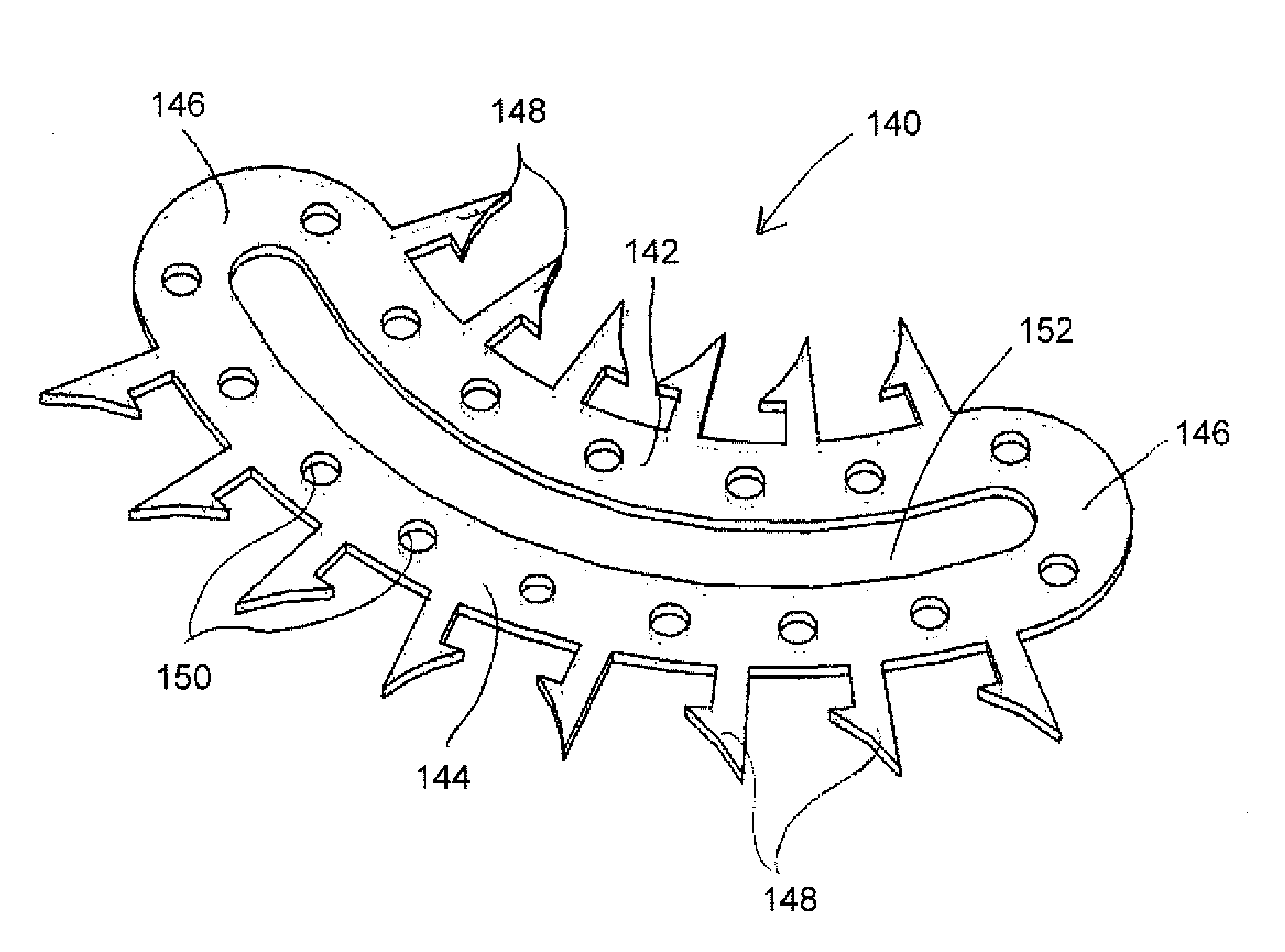
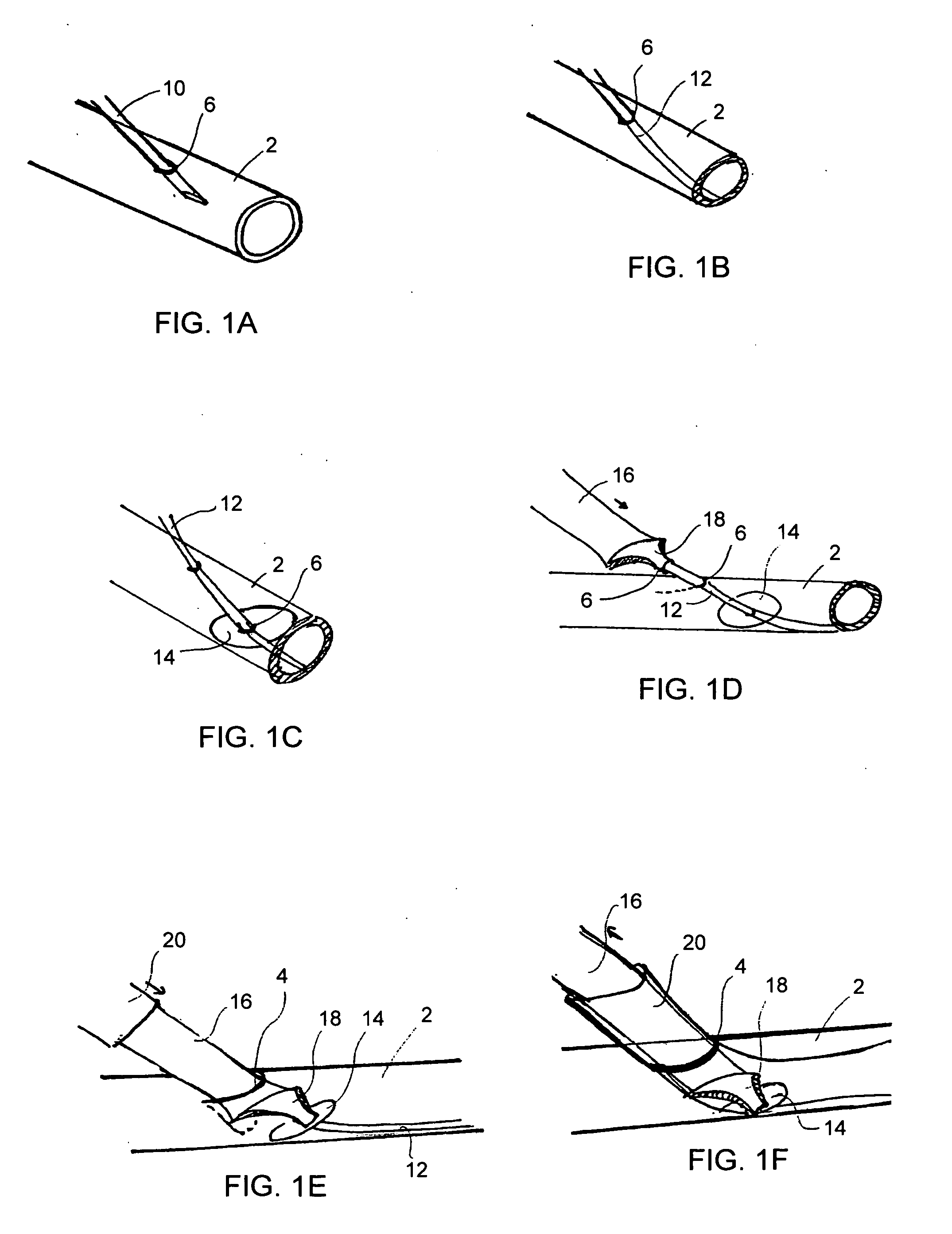
[0012]Certain variations of these implantable devices are configured to be implanted, fixed or placed subsequent to completion of the diagnostic or therapeutic procedure while others are configured to be implanted, fixed or placed prior to performing the procedure. Of the latter variety, certain of these devices are placed and affixed to the target vessel or tissue even prior to forming an incision within the vessel or tissue. Still yet, certain embodiments of the pre-incision implants allow for the precise formation of an incision which forms the access aperture and subsequent control of the opening (for the passage of instruments and other devices there through) and closing (after the removal of all instrumentation) of the access aperture. More particularly, the pre-incision implants precisely define the location, shape, size and length of the aperture to be formed, allow for controlled formation and maintenance of that aperture, and allow for precise apposition of the edges of the vessel aperture for optimal sealing of the aperture incision.
[0013]The implantable devices may be fabricated of materials which have elastic characteristics, such as superelastic materials, that allow the device to be transitioned from / to a functional state to / from a lower-profile or compressed state and back again where the device, when in the lower-profile state, has at least one dimension that is less than when in the other state. When in a lower-profile state, the device facilitates its percutaneous delivery to a target tissue site, and can subsequently be released or expanded to the functional state upon positioning at the target site. When in the functional or expanded state, the device allows for the controlled opening and closing of the incision.
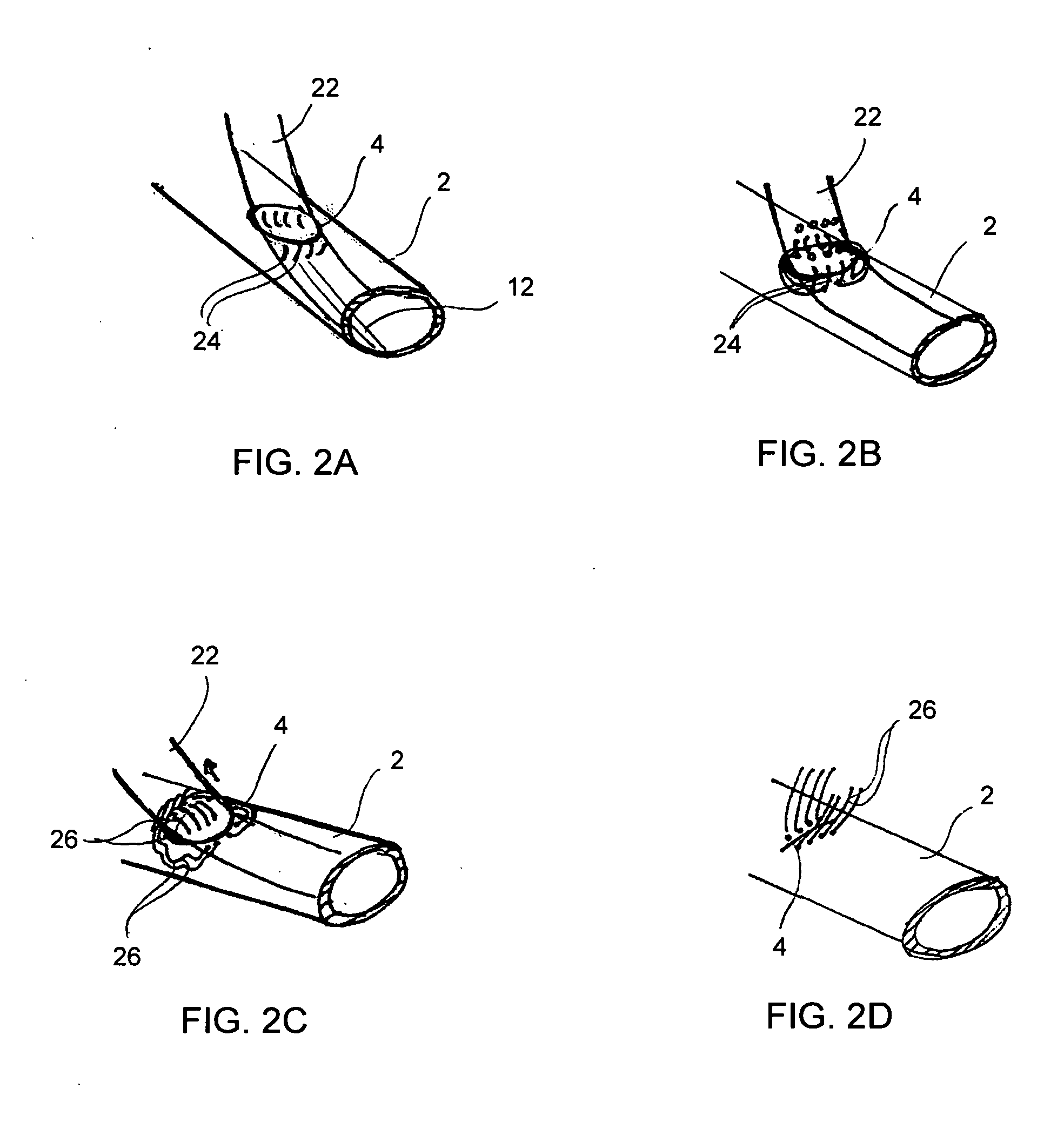
[0016]The present invention also provides methods, which include using the subject implantable devices and / or the subject systems to form access apertures within tissue structures and / or for closing those apertures. Certain of these methods include facilitating the performance of percutaneous or endovascular procedures through a controlled tissue opening.

Problems solved by technology

While some surgeons have used existing vascular closure devices to close large arteriotomy sites, such has proven difficult, unreliable, and therefore not widely-adopted.
Not only is there a lack of effective percutaneous devices and methodologies for the closure of large diameter vessel openings, the same can be said for the creation of such large openings.
While nominally traumatic when used to create smaller vessel openings, larger dilators can significantly traumatize the skin and the vessel tissue.
This potentially causes injury and delamination of tissue layers in the wound tract.
Furthermore, the stretching and tearing of the vessel wall results in an opening which is not uniform with an unpredictable shape and size.
Moreover, the edges of the vessel opening can become friable and misshapen, making subsequent closure that much more difficult.
Specifically, without the ability to provide a clean edge-to-edge alignment when closing a vessel opening, hemostasis is made difficult and endothelial and intimal growth between the vessel edges is impaired, thereby negatively affecting the wound's ability to heal properly.

Method used


Image

  • Devices and methods for the controlled formation and closure of vascular openings
  • Devices and methods for the controlled formation and closure of vascular openings
  • Devices and methods for the controlled formation and closure of vascular openings

Examples


embodiment 250

[0061]FIGS. 7A and 7B illustrate an exemplary frame embodiment 250 of the present invention having an inner frame 252 and outer frame 254. FIG. 7A shows frame 250 in the configuration which it would have when operatively engaged with the outer surface of a vessel wall upon securement thereto. FIG. 7B illustrates the relative movement of the inner and outer frame structures in operative use. In particular, inner frame 252 has been pushed downward out of plane relative to outer frame 254; however, the inner frame may pulled upward relative to the outer frame and / or the outer frame may be moved in either direction relative to the inner frame.

[0062]The extent to which the inner frame member (and / or outer frame member), and thus the tissue flap, can be opened or spaced from the outer frame or surface of the vessel wall to which a subject frame is positioned depends at least in part on the physical properties of the material used to make the frame and the thickness of the frame. Typically...

embodiment 310

[0067]With certain embodiments of the implantable frames, a stopper mechanism or backstop may not be necessary in order to safely place and secure the frames at the implant site. For example, the frame embodiment 310 of FIGS. 6L and 6L′ does not require use of a stopper within the vessel interior. As illustrated, in FIGS. 10A-10C, frame 310 is delivered and positioned on and parallel to a surface of a blood vessel 318. Without a stopper mechanism, as the coil screws 316 are rotated into vessel wall 318, vessel wall 318 is initially compressed thereby closing off normal blood flow (see FIG. 10B). Continued rotation and downward pressure on coil screws 316 causes them to penetrate vessel wall 318, possibly as well as the opposing vessel wall 318a, depending on their effective length. In any case, the lengths of the coil screws 316 are such that that any penetration into the opposite wall would be nominal with the penetrated length being easily retracted from the opposite tissue wall b...


PUM


Abstract

The present invention includes systems, devices and methods for percutaneously forming an aperture within a tissue structure or vessel and closing the aperture in a manner which optimizes hemostasis and healing. The invention in one aspect includes implantable devices which are used to seal the tissue aperture upon closure of the aperture after a percutaneous or endovascular procedure.

Description

CROSS REFERENCE[0001]This filing claims the benefit of provisional patent application Ser. No. ______, entitled “Device for Controlled Opening of Vessels” filed May 25, 2005, the entirety of which is incorporated by reference.FIELD OF THE INVENTION[0002]The present invention relates to the percutaneous formation and closure of vascular openings. The present invention is particularly advantageous for forming and closing large-diameter vascular openings.BACKGROUND OF THE INVENTION[0003]Access to patient blood vessels is necessary for a wide variety of diagnostic and therapeutic purposes. For example, intravascular catheters are introduced to both the arterial vasculature and the venous vasculature, typically using either surgical cut down techniques or percutaneous introduction techniques in which an opening is created in the wall of a vessel situated relatively close to the skin surface.[0004]The continued popularization of minimally invasive and endovascular procedures and the adven...

Claims


Application Information

Patent Timeline
IPC IPC(8): A61B17/10A61B17/08
CPCA61B17/0057A61B17/064A61B17/068A61B2017/0649A61B2017/00663A61B2017/00867A61B2017/0641A61B2017/00637
Inventor BENJAMIN, ELADGOBER, MICHAEL ELLIOTPEINKNAGURA, CARLASAFA, BAUBACKTHOMAS, JONATHAN MOULTON
Owner THE BOARD OF TRUSTEES OF THE LELAND STANFORD JUNIOR UNIV
Learn More