Systems and methods to analyze multiplexed bead-based assays using backscattered light

a technology of backscattered light and analysis methods, applied in the field of microscopy, can solve the problems of increasing processing time proportionally, increasing computational costs, and prone to optical alignment problems in imaging systems with multiple sensors to image the same fov, so as to reduce imaging and analysis time, improve image quality, and reduce the number of images

Active Publication Date: 2008-10-09
NORTHWESTERN UNIV
View PDF20 Cites 12 Cited by
  • Summary
  • Abstract
  • Description
  • Claims
  • Application Information

AI Technical Summary

Benefits of technology

[0008]One embodiment of the invention relates to an epifluorescence microscope based optical system equipped with a tunable filter to localize microspheres in bead-based assays based on a back-scattered light image (also known as reflected light image) and to read emission spectra from the microspheres. A common optical path for the reflected light and luminescent emission from the beads in conjunction with a tunable filter negates the requirement of an additional sensor employed in existing technologies for localizing microspheres based on light scatter measurements.
[0009]In addition to locating and identifying the luminescence emission of the beads, the present systems may be used to identify and quantify target molecules immobilized on the beads. In this embodiment, the systems may use target molecules having attached labels, wherein the labels emit a luminescent signal when excited by the excitation radiation. The labels may be, but are not limited to, fluorescent labels, phosphor labels, and / or scattering labels. Specific examples of labels that may be associated with the target molecules include fluorescent dyes, quantum dots and colloidal gold. The labels desirably have excitation wavelengths as well as emission wavelengths that overlap with the excitation wavelength band of the luminescently coded beads. For example, the excitation and emission wavelengths of the labels may overlap with the excitation wavelength band for the QDs used for labeling polymer microspheres. In this embodiment of the method, images acquired at emission wavelengths of the target molecules can be utilized for localizing the microspheres as well as for identifying and quantifying the target molecules. This reduces the number of images per FOV required for analysis, thereby reducing the imaging and analysis time, The system also allows for the utilization of uncoded or blank microbeads as assay platforms. An image analysis algorithm, which compensates for any small drift of microspheres in the array, may optionally be employed to improve the image.

Problems solved by technology

The analysis is computationally intensive and the processing time increases proportionally with the increase in number of colors (i.e., wavelengths).
However, in all of these methods, the imaging apparatus requires a different sensor (such as a photomultiplier tube) to measure the forward scatter signal.
An imaging system with multiple sensors to image the same FOV is more susceptible to optical alignment problems and prone to errors in the signal measurements.

Method used


Image

  • Systems and methods to analyze multiplexed bead-based assays using backscattered light
  • Systems and methods to analyze multiplexed bead-based assays using backscattered light
  • Systems and methods to analyze multiplexed bead-based assays using backscattered light

Examples


Embodiment Construction

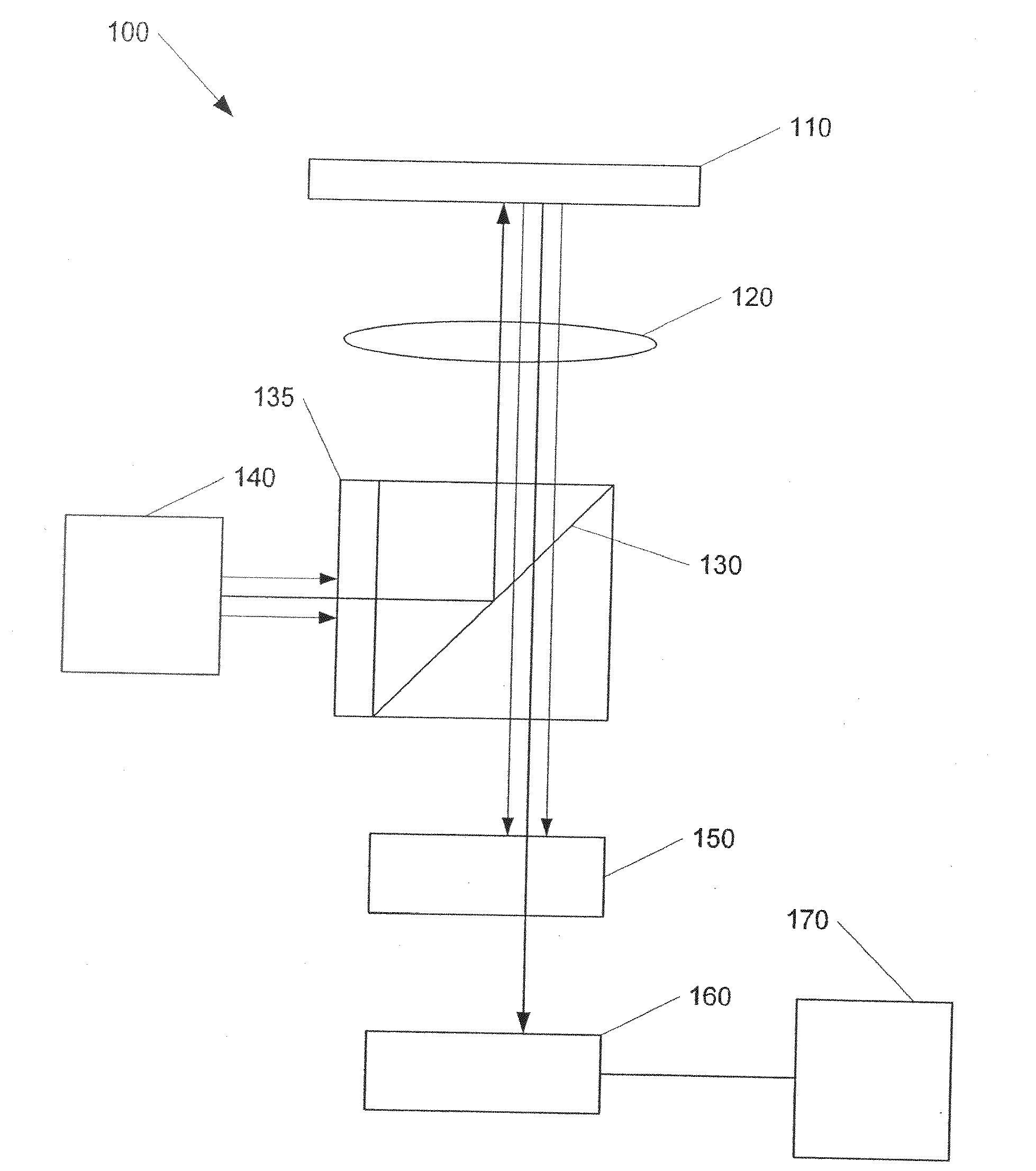
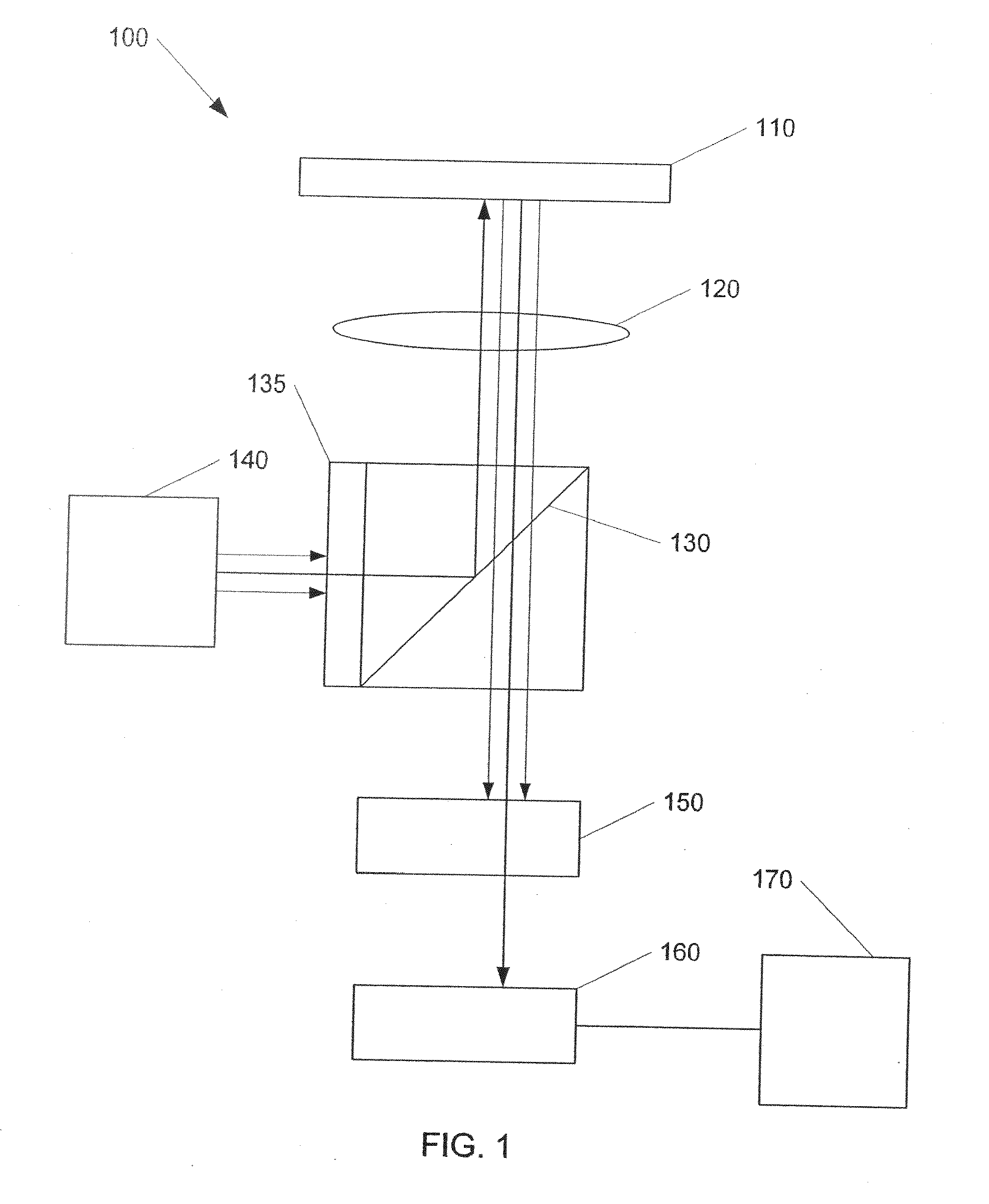
[0015]To overcome the limitations of a multisensor imaging system, novel methods and systems have been developed in which back-scattered light (also known as ‘reflected light’) from beads in a sample in an FOV is utilized to localize the beads and emitted luminescence signal is read from the corresponding locations in the images at various emission wavelengths to determine the spectral code for each bead. The system, which does not require any additional sensor for measuring the reflected light signal, tunes a tunable filter to the excitation wavelength in order to detect back-scattered light, similar to detecting the emitted luminescence signal when the tuning filter is tuned to different emission wavelengths. The system helps to eliminate any calibration errors that occur due to misalignment problems in systems with multiple sensors. As used herein, the term “tunable filter” refers to any device that is capable of being tuned to selectively transmit radiation of different waveleng...


PUM

PropertyMeasurementUnit
wavelengthsaaaaaaaaaa
diametersaaaaaaaaaa

Abstract

This invention relates to a system and method related to an epifluorescence microscope based optical system equipped with a tunable filter to localize microspheres in bead-based assays based on a back-scattered light (also known as reflected light) image. A common optical path for reflected and emitted luminescence in conjunction with a tunable filter negates the requirement of an additional sensor employed in existing technologies for localizing microspheres based on light scatter measurements.

Description

CROSS-REFERENCE TO RELATED APPLICATIONS[0001]This application claims the benefit of U.S. Provisional Patent Application Ser. No. 60 / 833,581 filed Jul. 27, 2006, the entire disclosure of which is incorporated by reference.STATEMENT OF GOVERNMENT RIGHTS[0002]This invention was made with United States government support awarded by the following agencies: National Institute of Biomedical Imaging and Bioengineering, Award Number ROlEB001418.FIELD OF THE INVENTION[0003]The present invention relates generally to the field of microscopy. More particularly, this invention relates to apparatus, systems and methods for conducting microscopy studies using bead-based assays.BACKGROUND OF THE INVENTION[0004]Systems that acquire images of luminescently-labeled microspheres have proven useful for assays. In some such systems, images of assayed microspheres are acquired at multiple emission wavelengths. The luminescent spectral code and fluorescence from each microsphere can be read by superimposing...

Claims


Application Information

Patent Timeline
Patent Type & Authority Applications(United States)
IPC IPC(8): G01N21/31
CPCG01N21/6428G01N2021/6421
Inventor KELSO, DAVID M.MATHUR, ABHISHEK
Owner NORTHWESTERN UNIV
Why Eureka
Try Eureka