Non iterative shimming in magnetic resonance imaging in the presence of high LIPID levels

a magnetic resonance imaging and high lipid level technology, applied in the field of magnetic resonance imaging, can solve the problems of limiting the accuracy of the bsub>0 /sub>map obtained, the number and geometry of the shim coil available, and the inhomogeneity which is more difficult to detect and correct, so as to extend the availability of methods

Inactive Publication Date: 2008-10-23
ALBERT EINSTEIN COLLEGE OF MEDICINE OF YESHIVA UNIV
View PDF3 Cites 22 Cited by
  • Summary
  • Abstract
  • Description
  • Claims
  • Application Information

AI Technical Summary

Benefits of technology

[0014]If the relative lipid / water ratio is known, the contribution solely due to B0 inhomogeneity can be calculated for arbitrary B0 evolution times, including time intervals when they are exactly opposite in phase. Thus once the relative contributions of the water and lipid resonances have been determined, the phase contribution of each can be determined and the effect of the local B0 field can be calculated. Accordingly, an object of the present invention is to extend the availability of methods used in MR imaging in the brain to other organs where lipids are in high content and can exceed the water contribution (e.g. bone marrow from the human leg). The present invention describes a shim method for shimming an entire slice of the leg (including bone marrow and subcutaneous fat) and acquiring a whole slice spectroscopic image of the human leg.

Problems solved by technology

However, the object being imaged also has a significant effect on magnetic field homogeneity which can be more difficult to detect and correct.
Furthermore, the ability of shim coils to correct even accurately and precisely known inhomogeneity is limited by the number and geometry of the shim coils available.
However, the use of a single relatively short time interval limits the accuracy of the B0 maps obtained (2).
Although integral multiples of 1 / Δv can be used to increase the accuracy, this increases the probability that aliasing will occur and the severity of any aliasing that does occur.
These inaccuracies then result in errors in the calculated shims, necessitating additional iterations or in some cases incorrect solutions.
Thus, spectroscopic imaging studies outside the brain, which sample large regions of interest (ROIs), continue to be limited by the inability to simultaneously shim large regions automatically (15).
Although Dixon imaging is effective at distinguishing between water and lipid resonances, Dixon images do not themselves provide an accurate map of B0 inhomogeneity.

Method used


Image

  • Non iterative shimming in magnetic resonance imaging in the presence of high LIPID levels
  • Non iterative shimming in magnetic resonance imaging in the presence of high LIPID levels
  • Non iterative shimming in magnetic resonance imaging in the presence of high LIPID levels

Examples


Embodiment Construction

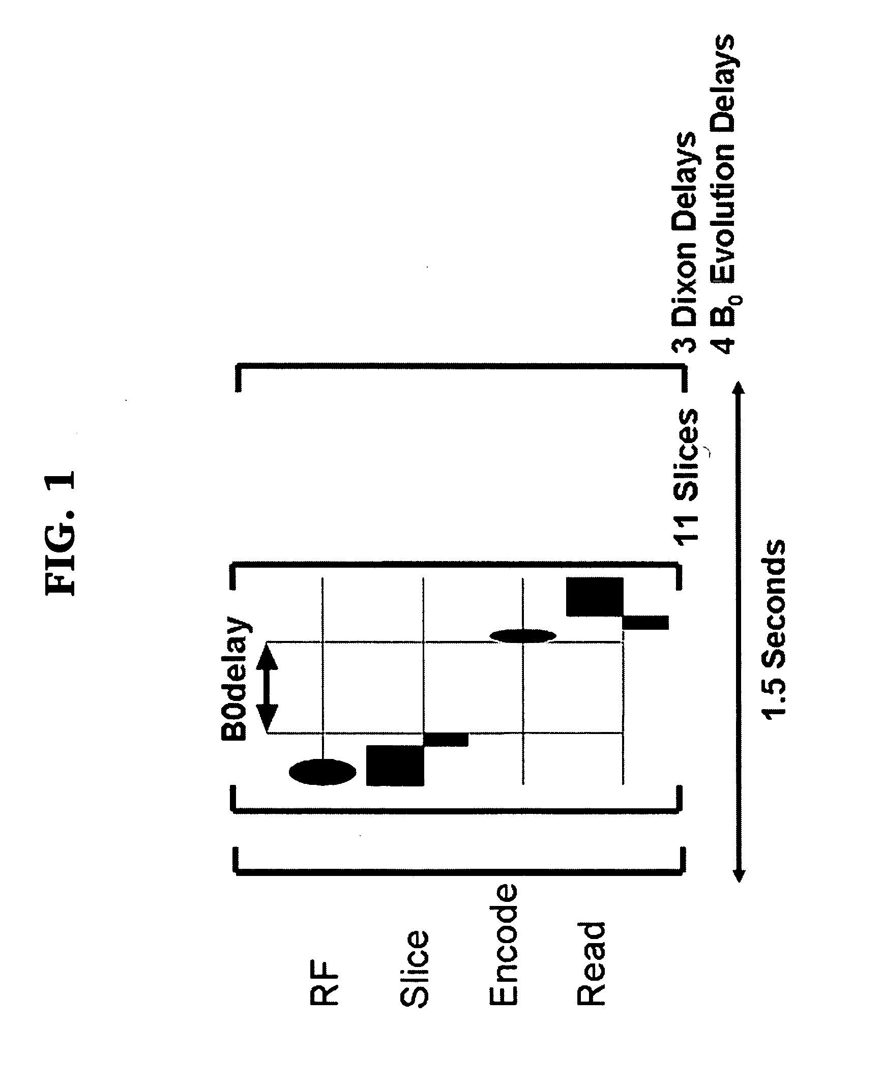
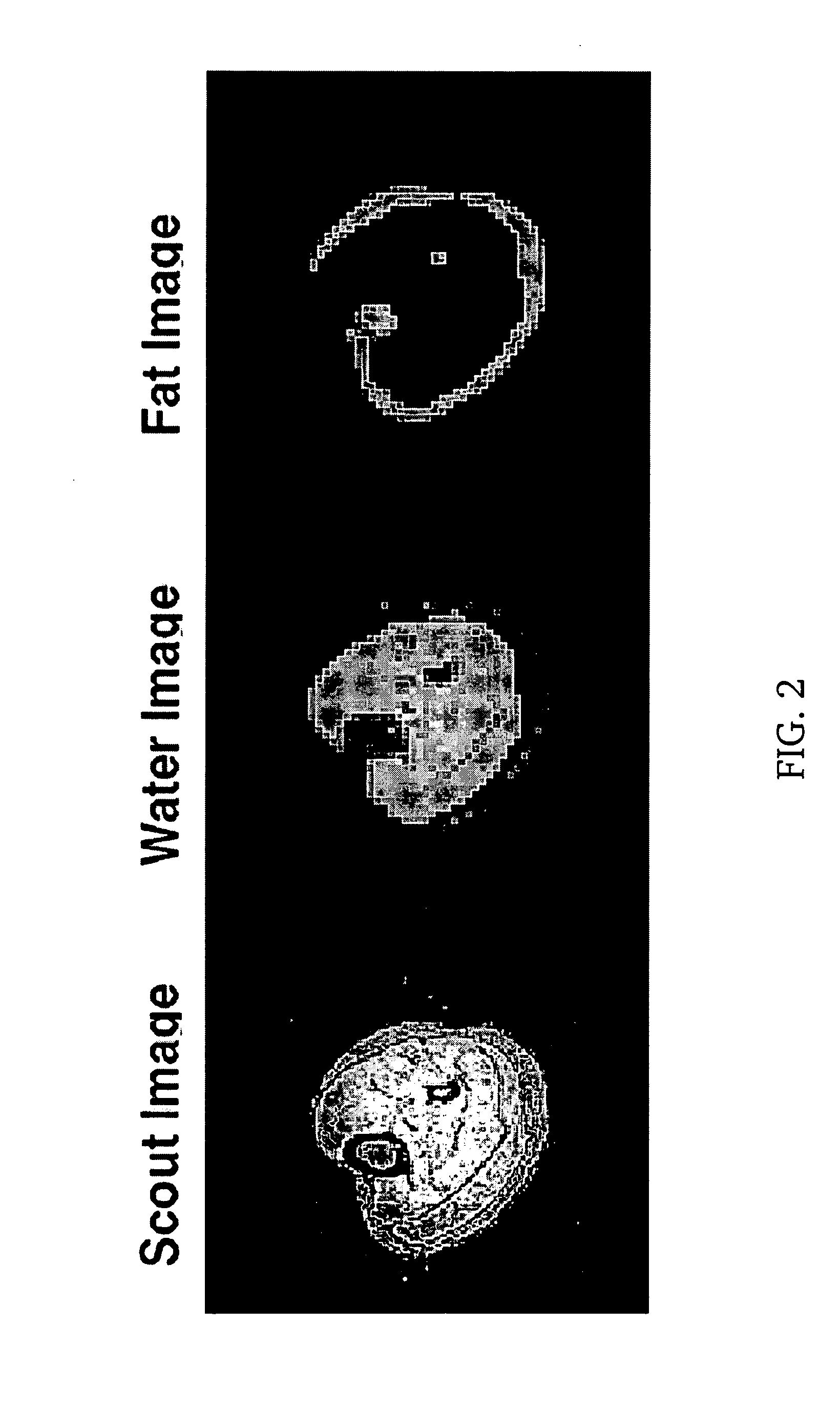
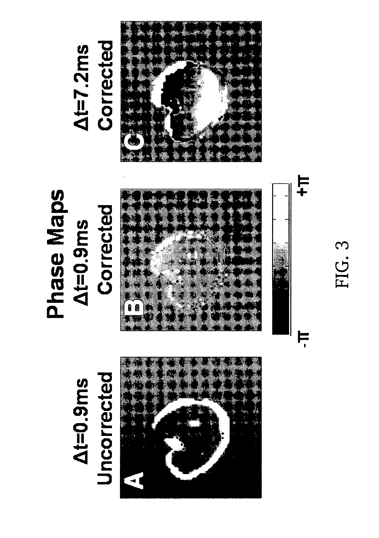
[0032]The invention provides a method for mapping the inhomogeneity of a magnetic field within an object in an MRI system, where the object contains a first and a second material, comprising the steps of:[0033]a) acquiring a plurality of images with the MRI system at a corresponding plurality of evolution times,[0034]b) calculating the phase contribution to the plurality of images due to the second material,[0035]c) removing said contribution from each of the plurality of images to obtain a phase map reflective of the inhomogeneity of the magnetic field,[0036]d) unwrapping aliasing present in the phase maps of images having relatively longer evolution times by reference to phase maps of images having relatively shorter evolution times, and[0037]e) generating a single map of magnetic field inhomogeneity from the unwrapped results.

[0038]Preferably, the first material is water and the second material has a Larmor frequency different from that of water. Preferably, the second material i...


PUM


Abstract

For the brain, a variety of automated non-iterative shimming methods using phase evolution derived B0 maps have been reported. These methods assume that there is only a single chemical species contributing to the image. Although true in the brain, lipid contributions from skin, bone marrow and structural fat, may approach or exceed the concentration of water in other organs. In these instances, standard B0 mapping methods cannot be used due to contributions arising from the lipids. To overcome these limitations the present invention discloses a multi-point B0 mapping method combined with Dixon imaging to provide fully automated shimming of the human calf.

Description

CROSS-REFERENCE TO RELATED APPLICATION[0001]This application claims the benefit of U.S. Provisional Patent Application Ser. No. 60 / 925,626, filed Apr. 20, 2007, the contents of which are incorporated by reference into the subject application.STATEMENT OF GOVERNMENT SUPPORT[0002]The invention disclosed herein was made with U.S. Government support under National Institutes of Health (NIH) grant numbers EB000473 and EB001748. Accordingly, the U.S. Government has certain rights in this invention.FIELD OF THE INVENTION[0003]The present invention relates to magnetic resonance (MR) imaging and more particularly to a method for mapping and reducing magnetic field variations when an object is in an imaging magnetic field.BACKGROUND OF THE INVENTION[0004]Throughout this application various publications are referred to in parenthesis. Full citations for these references may be found at the end of the specification immediately preceding the claims. The disclosures of these publications are here...

Claims


Application Information

Patent Timeline
Patent Type & Authority Applications(United States)
IPC IPC(8): G01R33/56
CPCG01R33/243G01R33/3875G01R33/4828G01R33/56563
Inventor HETHERINGTON, HOBY P.PAN, JULLIE W.
Owner ALBERT EINSTEIN COLLEGE OF MEDICINE OF YESHIVA UNIV
Why Eureka
Try Eureka