
Relative value summary perfusion map

a perfusion map and relative value technology, applied in the field of computed tomography (ct) perfusion imaging, can solve the problems of sudden loss of neurological function, irreversible damage of cells, and inability to accurately predict the perfusion rate,

Inactive Publication Date: 2008-10-23
BRUMMETT DAVID P
View PDF16 Cites 43 Cited by
  • Summary
  • Abstract
  • Description
  • Claims
  • Application Information

AI Technical Summary

Benefits of technology

[0015]In another embodiment, the step of using relative difference values to generate the composite includes a step of classifying the damaged tissues, e.g., ischemia, decreased perfusion without substantial cell injury, penumbra, infarct) into different perfusion states utilizing threshold values applied to each of the parameters measured (e.g., one or more of MTT, CBV, CCF, TTP).

Problems solved by technology

The condition typically causes a sudden loss of neurological function due to decreased perfusion of the brain with oxygen and glucose.
The decrease in perfusion can injure the affected brain cells—a condition referred to in the art as “ischemia.” If the decreased perfusion continues, the cell will be irreversibly damaged and eventually die.
But the decision to implement thrombolysis is not without risk.
The unnecessary implementation of thrombolysis where the damaged cells are beyond repair can lead to hemorrhage without the benefit of restoration of blood flow.
With sustained hypo-perfusion, however, the auto-regulatory mechanisms fail and the CBV decreases enough that tissue is no longer salvageable.
In these instances, the patient will likely have permanent injury.
Utilizing these parameters can be a practical problem because there is a tremendous amount of data to process in a very short period of time.
The longer the decreased perfusion occurs, the more the brain is damaged.
On the other hand, if therapy is incorrectly applied to brain that is not salvageable not only will brain cells not respond to the therapy, but the risk of therapy-induced brain hemorrhage increases.
The proposed methods, however, have limitations.
Using a value which is specific for gray or white matter can result in incorrect characterization of tissue type not encompassed by that value.
Further, reliance on absolute values also does not completely allow for variations that exist in these values even among normal patients, as there is a range of values that are considered normal.
Another problem is that the absolute value numbers generated can also vary when different operators of the CT scanner and post-processing equipment are performing the study (3, 4, 5).
Because of this, the user of the Wintermark technique may not be as accurate in making emergency treatment decisions.
Because the data images are not at all interrelated, the parameters cannot be weighted with respect to their particular strengths or weaknesses when displaying the perfusion data.
All of this causes the individual using this data to lose valuable time when evaluating a patient.
But this will only be executed if both of the location of interest and the substantially symmetrically opposite location are abnormal.

Method used


Image

  • Relative value summary perfusion map
  • Relative value summary perfusion map
  • Relative value summary perfusion map

Examples


Embodiment Construction

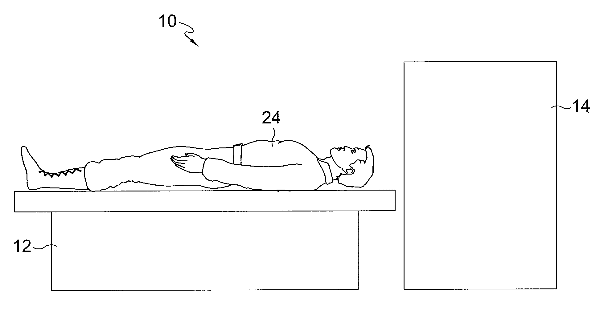
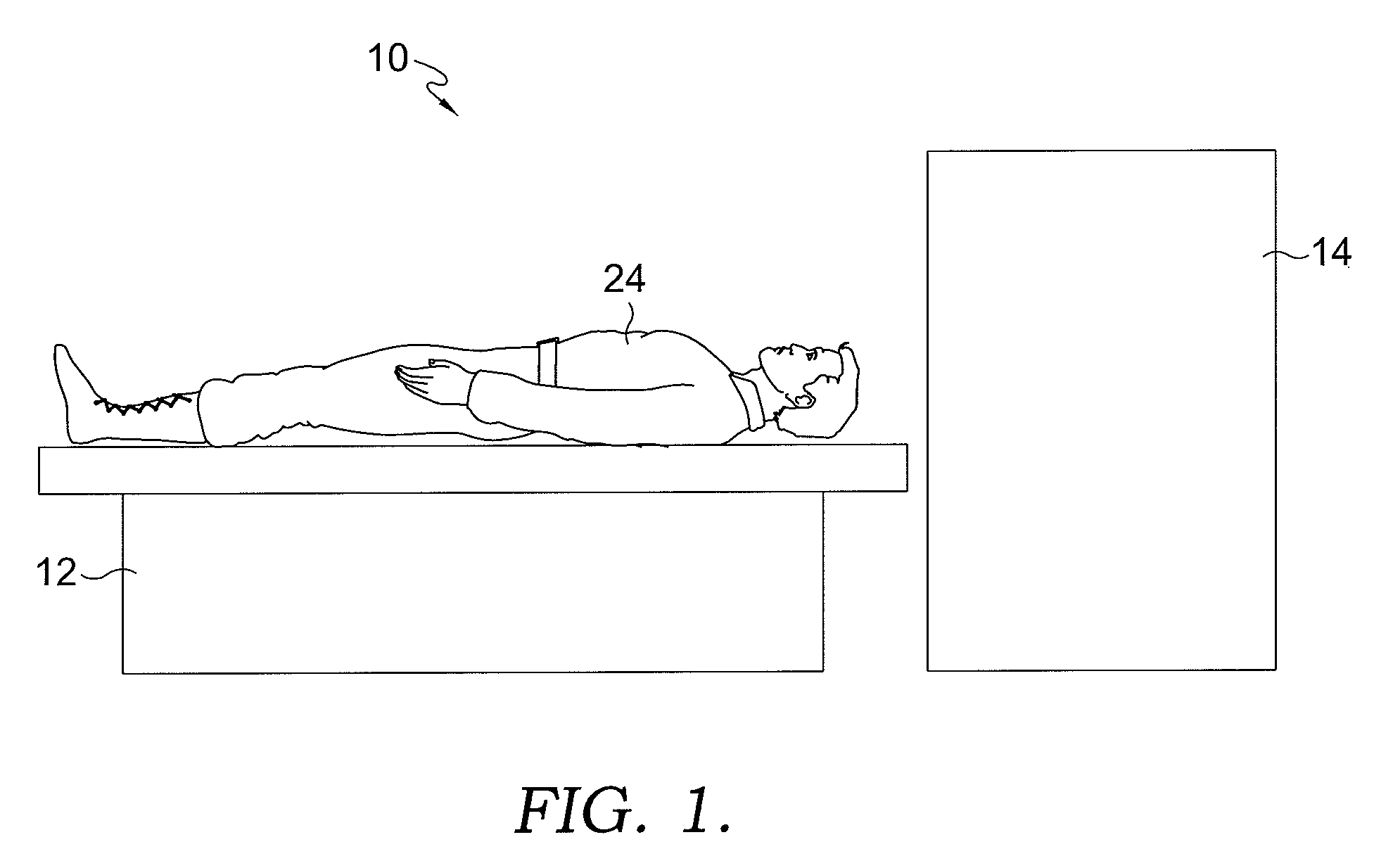
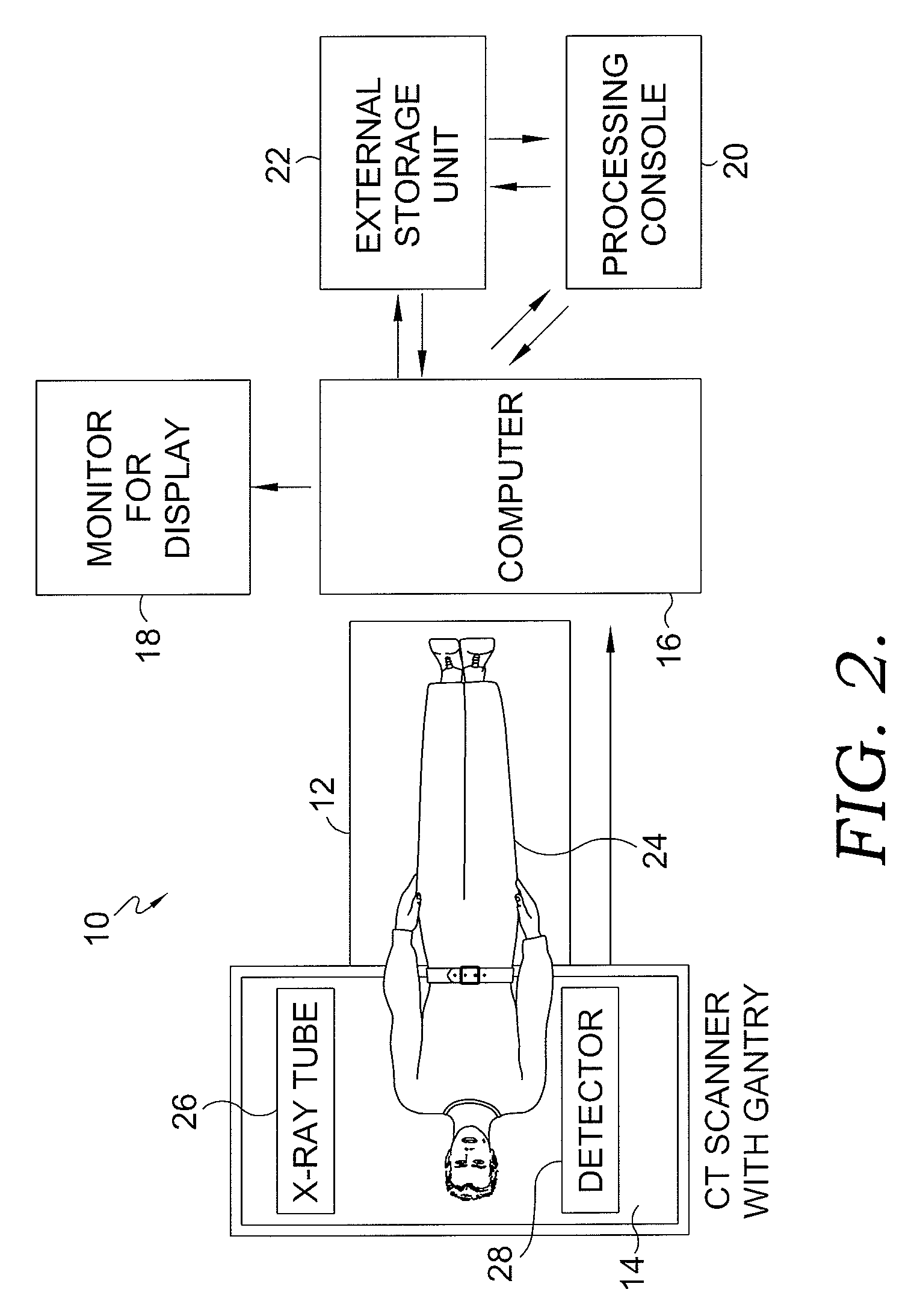
[0030]The disclosed invention relates to a system and a method for generating map and summary values that will simply review of the CT perfusion data. The map created will primarily utilize relative difference in perfusion among different brain regions and will be displayed as a composite image displaying a plurality of parameters at once. The diagnosing and treating physicians can then more quickly and accurately evaluate the patient's condition to determine whether to initiate or withhold thrombolytic therapy. The map can also provide general prognostic information. Because the process enables quantifying brain ischemia in a summary fashion, deciding which therapy option to use on the patient is accomplished more easily and faster. Further, the process is able to provide basic prognostic information regarding the degree of current brain injury and amount of injury that can exist if adequate perfusion is not restored with more accuracy than can be accomplished with the prior art me...


PUM


Abstract

Disclosed is a system and method for diagnosing a perfusion condition in the human brain. The process used includes measuring parameter values at a location of interest in one hemisphere and determining relative differences between those measured values against values measured at a substantially symmetrically opposite location in the other hemisphere. These relative differences are used to generate a composite output which is displayed using different colors. Each color is reflective of a different tissue state, e.g., ischemic core, penumbra, etc. If the substantially symmetrically opposite location is abnormal, and thus, is not a good reference, an alternative reference location which is not substantially symmetrically opposite the location of interest is chosen, and a relative difference map generated. Alternatively, an absolute value map can be generated.

Description

BACKGROUND OF THE INVENTION[0001]1. Field of the Invention[0002]The invention relates generally to the field of using computed tomography (CT) for perfusion imaging. More specifically, the invention relates to a system and a method for generating map and summary values for the purpose of enabling a user to more easily and quickly review of the CT perfusion data.[0003]2. Description of the Related Art[0004]Stroke is one of the leading causes of death and disability. The condition typically causes a sudden loss of neurological function due to decreased perfusion of the brain with oxygen and glucose. The decrease in perfusion can injure the affected brain cells—a condition referred to in the art as “ischemia.” If the decreased perfusion continues, the cell will be irreversibly damaged and eventually die. This is called infarction or stroke. The group of cells that have been damaged beyond the point of recovery have also been referred to as the ischemic core. If the decrease in perfusio...

Claims


Application Information

Patent Timeline
Patent Type & Authority Applications(United States)
IPC IPC(8): A61B6/00A61B5/05
CPCA61B5/0275A61B6/481A61B6/507A61B6/504A61B6/501A61B5/4064
Inventor BRUMMETT, DAVID P.
Owner BRUMMETT DAVID P
Learn More