Algorithms for selecting mass density candidates from digital mammograms

a technology of mass density and algorithm, applied in the field of medical imaging analysis, can solve the problem of fast processing time and achieve the effect of fast processing time and better selection sensitivity

Active Publication Date: 2008-10-30
THREE PALM SOFTWARE
View PDF6 Cites 23 Cited by
  • Summary
  • Abstract
  • Description
  • Claims
  • Application Information

AI Technical Summary

Benefits of technology

[0009]This invention makes use of both intensity and morphologic algorithms to process each image at a single gray-level to select the candidates. The detailed algorithm is shown in FIG. 2. Because both intensity and morphological information are used, the selection sensitivity is better than algorithms that use intensity alone. Since each image is processed at only one gray level, the processing time is fast. The typical time to generate around 25 candidates from one mammogram image is less than 500 ms, which is much faster than a comparable band-pass method, which typically takes more than 5 seconds for a single mammogram.

Problems solved by technology

Since each image is processed at only one gray level, the processing time is fast.

Method used


Image

  • Algorithms for selecting mass density candidates from digital mammograms
  • Algorithms for selecting mass density candidates from digital mammograms
  • Algorithms for selecting mass density candidates from digital mammograms

Examples


Embodiment Construction

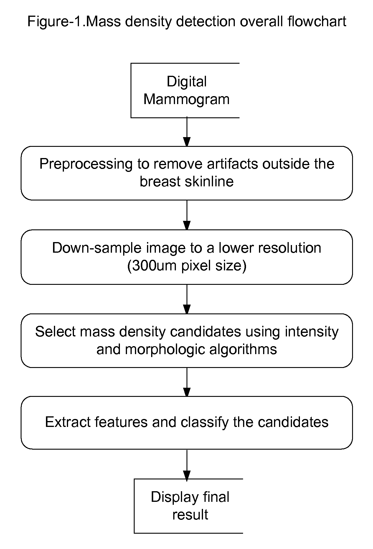
[0014]As shown in FIG. 1, the input to the mass candidate selection algorithm is a digital mammogram image, or a breast image from other modality. The image is preprocessed to remove artifacts outside the breast tissue. The image resolution of a digital mammogram is usually between 50 um to 100 um. The image therefore can be down-sampled to a lower resolution, i.e., 300 um, in order to improve processing speed without compromising processing quality. The algorithm to select mass density candidate uses this down-sampled image.

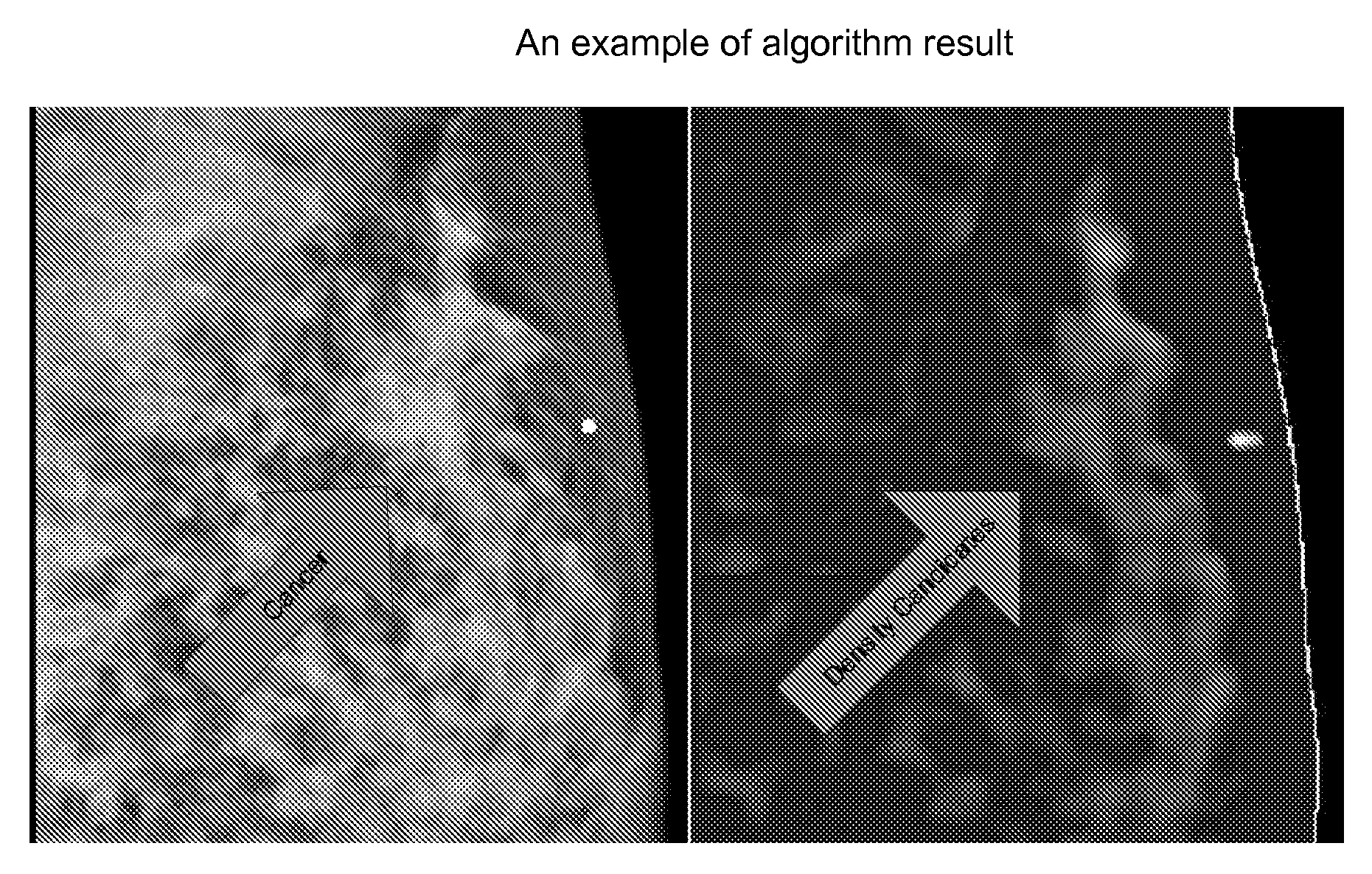
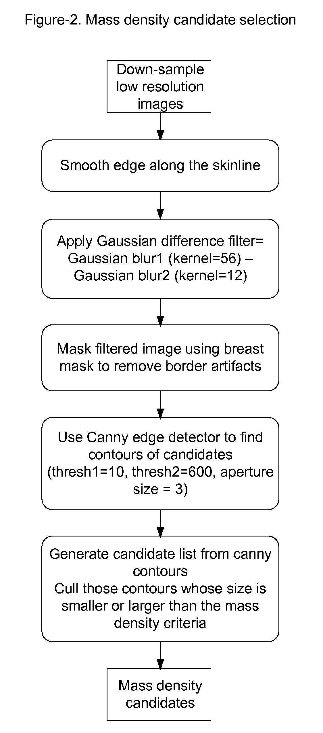
[0015]As shown in FIG. 2, the down-sampled image is smoothed along the edge of the skinline. A Gaussian difference filter is applied to the smoothed image (see FIG. 3 the original image and the Gaussian difference filtered image). The first Gaussian filter kernel size is selected as 56; and the second Gaussian filter kernel size is selected as 12. The filtered image is masked by breast mask to remove border artifacts. Next step is to use Canny edge detector to f...


PUM


Abstract

The present invention provides a method for selecting mass density candidates from mammograms for computer-aided lesion detection, review and diagnosis. The method has two steps: a Gaussian difference filter to enhance the intensity and a Canny detector to find potential mass density contours. For circumscribed masses, an additional Hough circle detector is used. This invention makes use of both intensity and morphology information and only processes each image at a single gray-level, so both sensitivity and processing time are improved. The selection algorithm can be also used to select mass candidates from ultrasound images, from 3D tomosynthesis mammography images and from breast MRI images.

Description

CROSS-REFERENCE TO RELATED APPLICATIONSU.S. Patent Documents [0001]1. U.S. Pat. No. 5,615,243 March 1997 Chang et al. “Identification of suspicious mass regions in mammograms”[0002]2. U.S. Pat. No. 5,832,103 November 1998 Giger et al. “Automated method and system for improved computerized detection and classification of masses in mammograms”[0003]3. U.S. Pat. No. 6,246,782 June 2001 Shapiro et al. “System for automated detection of cancerous masses in mammograms”STATEMENT REGARDING FEDERALLY SPONSORED RESEARCH OR DEVELOPMENT[0004]Not Applicable.REFERENCE TO SEQUENCE LISTING, A TABLE, OR A COMPUTER PROGRAM LISTING COMPACT DISC APPENDIX[0005]Not Applicable.BACKGROUND OF THE INVENTION[0006]The present invention relates generally to the field of medical imaging analysis. Particularly, the present invention relates to a method and system for candidates selection of mass density from digital mammography images in conjunction with computer-aided detection, review and diagnosis (CAD) for ma...

Claims


Application Information

Patent Timeline
Patent Type & Authority Applications(United States)
IPC IPC(8): G06K9/00
CPCG06T7/0012G06T2207/10088G06T2207/10112G06T2207/10116G06T2207/10132G06T2207/30068
Inventor ZHANG, HEIDI DAOXIANHEFFERNAN, PATRICK BERNARD
Owner THREE PALM SOFTWARE
Why Eureka
Try Eureka