Eureka AIR delivers breakthrough ideas for toughest innovation challenges, trusted by R&D personnel around the world.

X-ray ct apparatus, myocardial perfusion information generating system, x-ray diagnostic method and myocardial perfusion information generating method

a technology of myocardial perfusion and computed tomography, which is applied in the field of x-ray ct (computed tomography) apparatus, myocardial perfusion information generating system, x-ray diagnostic method and myocardial perfusion information generating method, can solve the problem of increasing the x-ray dosage of the object, and achieve the effect of reducing the amount of contrast medium injection

Active Publication Date: 2008-11-06
TOSHIBA MEDICAL SYST CORP
View PDF2 Cites 17 Cited by
  • Summary
  • Abstract
  • Description
  • Claims
  • Application Information

AI Technical Summary

Benefits of technology

[0012]The present invention has been made in light of the conventional situations, and it is an object of the present invention to provide an X-ray CT apparatus, a myocardial perfusion information generating system, an X-ray diagnostic method and a myocardial perfusion information generating method which can acquire myocardial perfusion information useful for diagnosis in a shorter period while reducing the amount of injection of a contrast medium as to an object and dosage due to X-ray.
[0017]The X-ray CT apparatus, the myocardial perfusion information generating system, the X-ray diagnostic method and the myocardial perfusion information generating method as described above make it possible to acquire myocardial perfusion information useful for diagnosis in a shorter period while reducing the amount of injection of a contrast medium as to an object and dosage due to X-ray.

Problems solved by technology

However, normally, such perfusion imaging is not an isolated examination, but performed as part of a contrast examination.
Accordingly, there is a problem that it takes a long time for scanning of a myocardial perfusion image, which results in increase of X-ray dosages for the object.

Method used


Image

  • X-ray ct apparatus, myocardial perfusion information generating system, x-ray diagnostic method and myocardial perfusion information generating method
  • X-ray ct apparatus, myocardial perfusion information generating system, x-ray diagnostic method and myocardial perfusion information generating method
  • X-ray ct apparatus, myocardial perfusion information generating system, x-ray diagnostic method and myocardial perfusion information generating method

Examples


Embodiment Construction

[0029]An X-ray CT apparatus, a myocardial perfusion information generating system, an X-ray diagnostic method and a myocardial perfusion information generating method according to the present invention will now be described in further detail below with reference to embodiments in conjunction with the accompanying drawings.

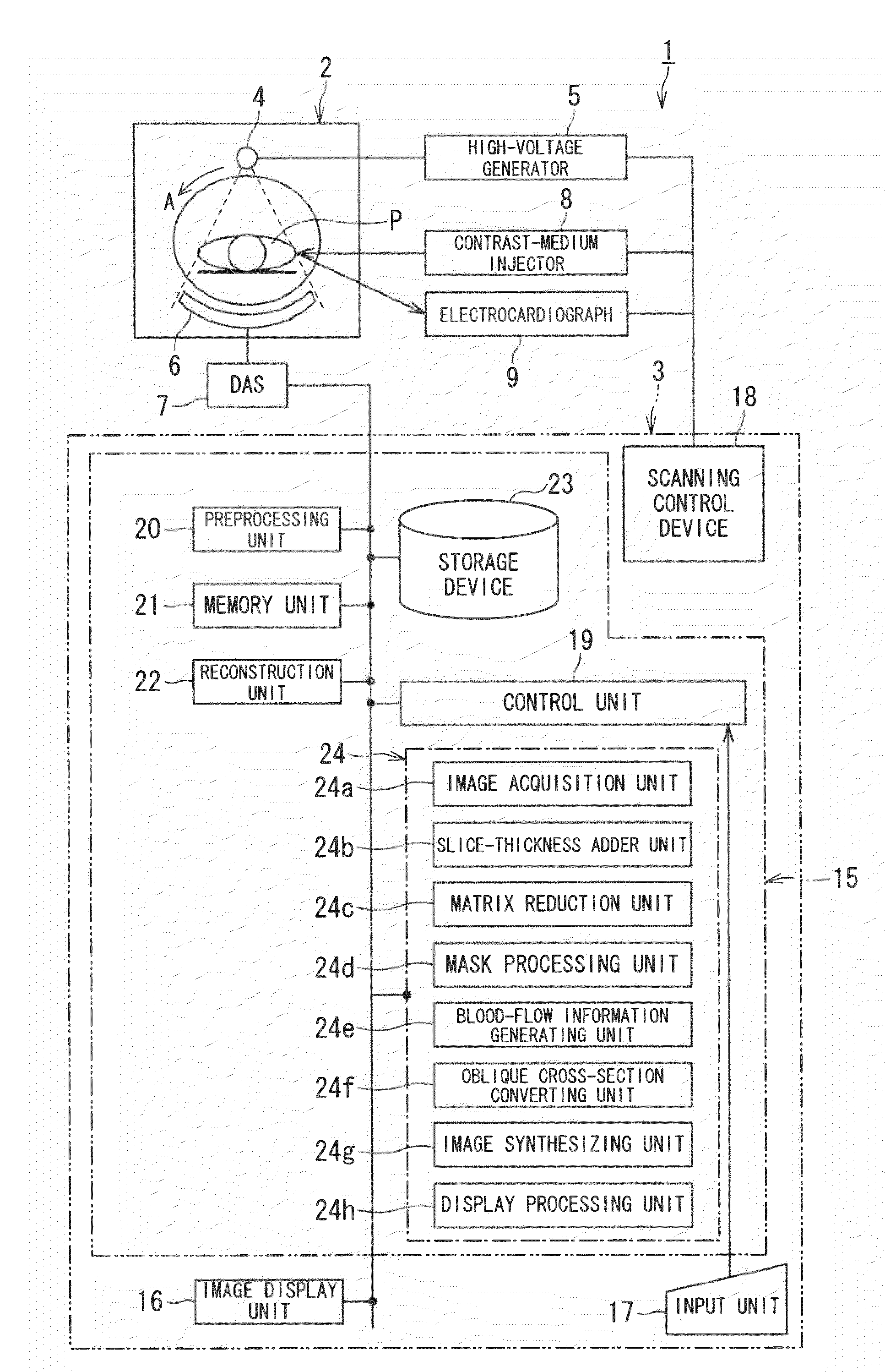
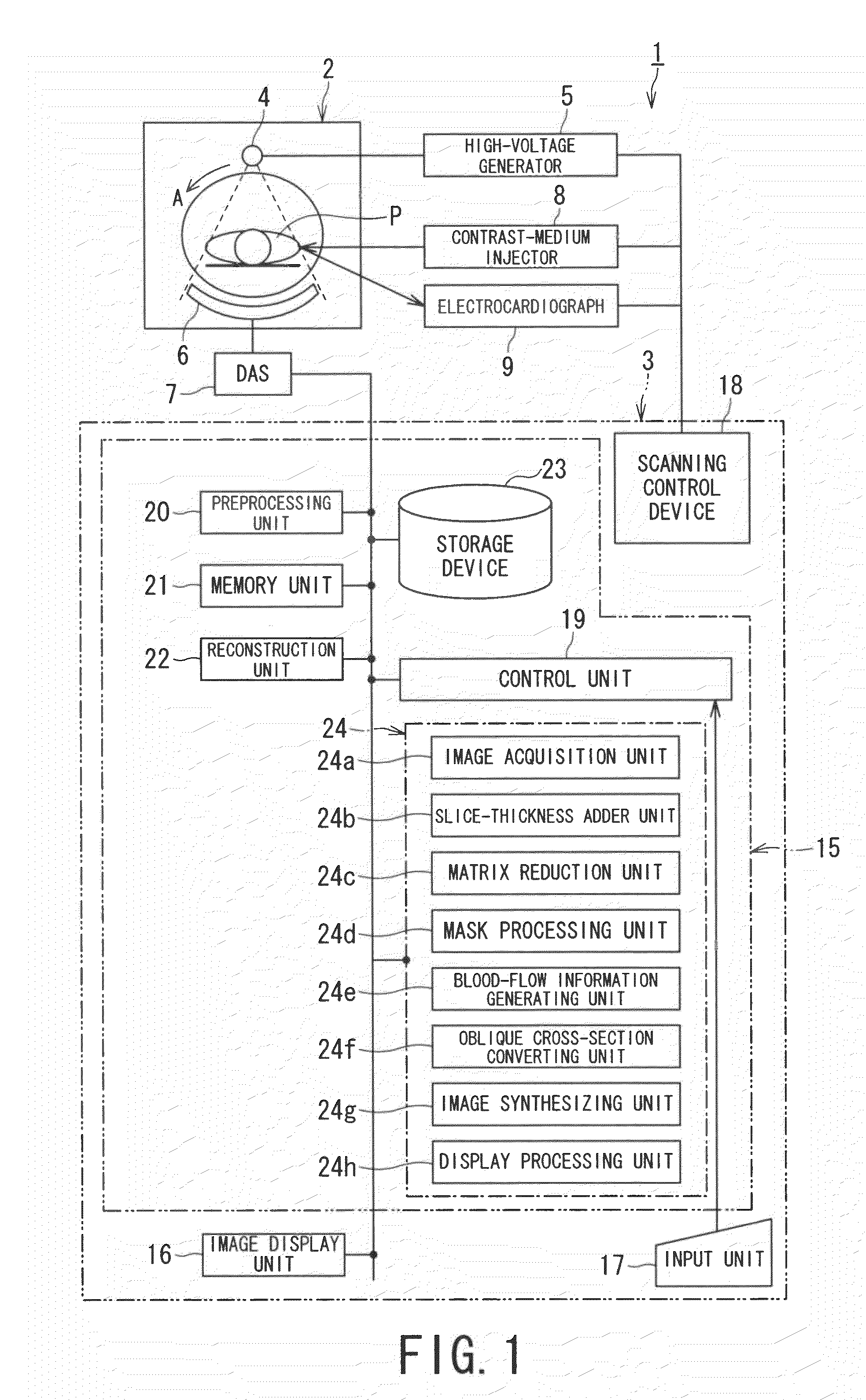
[0030]FIG. 1 is a configuration diagram illustrating an X-ray CT apparatus according to an embodiment of the present invention.

[0031]An X-ray CT apparatus 1 includes a gantry unit 2 and a computer device 3. The gantry unit 2 includes an X-ray tube 4, a high-voltage generator 5, an X-ray detector 6, a DAS (Data Acquisition System) 7, a contrast-medium injector 8, and an electrocardiograph 9. The X-ray tube 4 and the X-ray detector 6 are mounted at positions facing each other sandwiching an object P in an unshown rotating ring consecutively rotating at a high speed.

[0032]The X-ray CT apparatus 1 has a function to generate contrast CT image data of the object P under ...


PUM


Abstract

An X-ray CT apparatus has an image generating unit and a blood flow information acquiring unit. The image generating unit acquires projection data at rest and at non-rest from a myocardial part of an object into which contrast medium is injected continuously in a state in which each concentration of the contrast medium can be considered to be constant and reconstructs contrast CT image data at the rest and at the non-rest using the acquired projection data. The blood flow information acquiring unit obtains third blood flow information based in first blood flow information derived from the contrast CT image data at the rest and second blood flow information derived from the contrast CT image data at the non-rest.

Description

BACKGROUND OF THE INVENTION[0001]1. Field of the Invention[0002]The present invention relates to an X-ray CT (computed tomography) apparatus, a myocardial perfusion information generating system, an X-ray diagnostic method and a myocardial perfusion information generating method for generating myocardial perfusion information with continuously injecting a contrast medium.[0003]2. Description of the Related Art[0004]With a myocardial contrast examination using an X-ray CT apparatus, a contrast medium is injected into an object that is a patient, and contrast CT images are collected. Subsequently, moving images of coronary arteries, endocardial lumen wall, and so forth, are generated from the collected contrast CT images, and are employed for diagnosis.[0005]Also, a blood flow dynamic state (perfusion) examination of the myocardium and a perfusion examination regarding organs such as within a brain tissue have been performed using an X-ray CT apparatus. With these perfusion examinatio...

Claims


Application Information

Patent Timeline
IPC IPC(8): G06K9/78
CPCA61B5/0275A61B6/481A61B6/503A61B6/504A61B6/507
Inventor ICHIHARA, TAKASHI
Owner TOSHIBA MEDICAL SYST CORP
Why Eureka
Eureka Blog
Learn More