System and method to generate an illustration of a cardiac region of interest

a cardiac region and illustration technology, applied in the field of medical imaging, can solve the problems of difficult to identify or correlate a defect detected in the cardiac tissue or muscle of the heart, and difficult to correlate the acquired measurements relative to acquired anatomical information

Active Publication Date: 2008-11-06
GENERAL ELECTRIC CO
View PDF9 Cites 90 Cited by
  • Summary
  • Abstract
  • Description
  • Claims
  • Application Information

AI Technical Summary

Problems solved by technology

However, a drawback of the above-described representation is a difficulty to correlate the acquired measurements relative to acquired anatomical information (e.g., position of the coronary arteries) acquired in tomography datasets.
In particular, it is generally very difficult to identify or correlate a defect detected in the cardiac tissue or muscle of the heart with a problem occurring in one of the coronary arteries.

Method used


Image

  • System and method to generate an illustration of a cardiac region of interest
  • System and method to generate an illustration of a cardiac region of interest
  • System and method to generate an illustration of a cardiac region of interest

Examples


Embodiment Construction

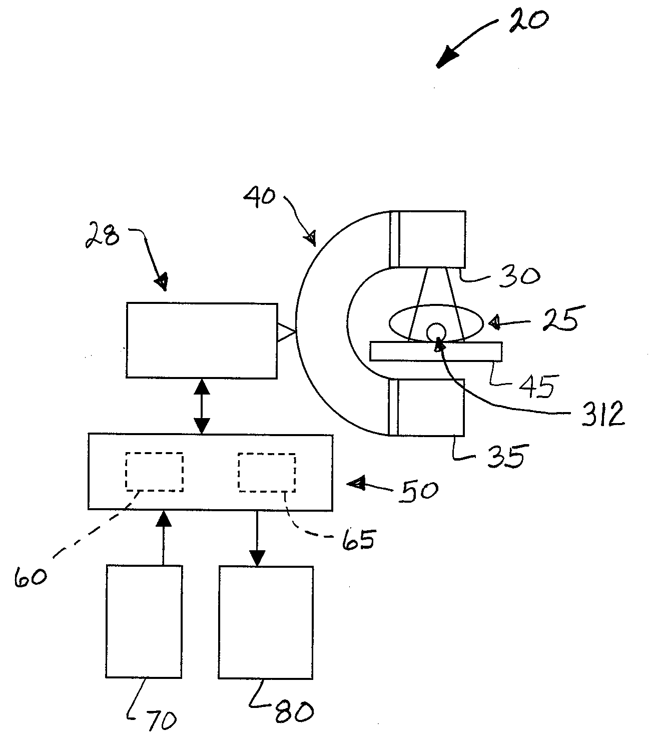
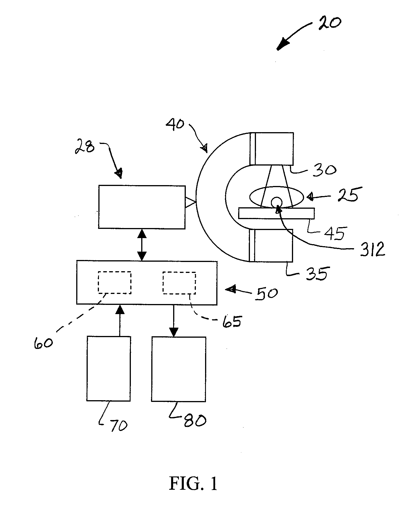
[0015]FIG. 1 shows an embodiment of a system 20 to generate a display of detected activity of a cardiac tissue of an imaged subject 25. The system 20 generally includes an imaging system 28 operable to acquire images of the imaged subject 25 from various positions. An embodiment of the imaging system 28 includes a radiation source 30 operable to emit radiation (e.g., X-rays) through an imaged subject 25 toward a detector 35. The system 20 also includes a gantry 40 in mobile support of both the source 30 and detector 35 in relation to a table 45 in support of the imaged subject 25. Yet, the type (e.g., radiological, computed tomography (CT), positron emission tomography (PET), magnetic resonance imaging (MRI), ultrasound, fluoroscopic, laparoscopic, etc.) of imaging system 28 can vary.

[0016]The system 20 also generally includes a controller 50 in communication (e.g., via direct wired links, fiber optics, wireless communications, etc.) with the source 30 and detector 35. The controlle...


PUM


Abstract

A system and method to generate an illustration of a cardiac region of interest of an imaged subject is provided. The method includes generating a three-dimensional model from a series of acquired images of the cardiac region of interest; measuring a series of values of at least one functional parameter of the cardiac region of interest; generating a map of a spatial relation of the plurality of values of the functional parameter in spatial relation to the three-dimensional model of the cardiac region of interest; generating a three-dimensional model of a vessel structure leading to the cardiac region of interest; generating an output image that includes combining the three-dimensional model of the cardiac region of interest, the map of the series of values of the functional parameter, and the three-dimensional model of the vessel structure in spatial relation to one another relative to a common coordinate system.

Description

BACKGROUND OF THE INVENTION[0001]The subject matter described herein generally relates to medical imaging, and more particularly to a system and method to generate images illustrative of a measured parameters of a region of interest (e.g., cardiac tissue of the heart) of an imaged subject.[0002]There exists a conventional multiphase computerized tomography analysis technique to detect a potential defect in the heart (e.g., the myocardium) of a patient. A certain known representation produced by such a conventional analysis is shown as a plurality of concentric circles. Typically, all the measurements of the above-described technique are shown in a single, two-dimensional illustration of the heart muscle under analysis.[0003]However, a drawback of the above-described representation is a difficulty to correlate the acquired measurements relative to acquired anatomical information (e.g., position of the coronary arteries) acquired in tomography datasets.[0004]In particular, it is gener...

Claims


Application Information

Patent Timeline
Patent Type & Authority Applications(United States)
IPC IPC(8): A61B6/00G06T19/00
CPCA61B6/032A61B6/463A61B6/503A61B6/504G06T5/50G06T7/0012G06T7/0083G06T2207/10081G06T2207/20221G06T2207/30048G06T2207/30101G06T19/20G06T2210/41G06T2219/2004G06T7/12
Inventor DESCHAMPS, THOMASKNOPLIOCH, JEROME F.LAUNAY, LAURENTNAY, MARIA-MAGDALENA
Owner GENERAL ELECTRIC CO
Why Eureka
Try Eureka