Micromachined membrane filter device for a glaucoma implant and method for making the same

a filter device and micro-machine technology, applied in the field of medical devices, can solve the problems of high intraocular pressure of glaucoma, high labor intensity of surgeons, and failure of procedures, and achieve the effect of limiting the ingress of microorganisms

Inactive Publication Date: 2008-11-13
BECTON DICKINSON & CO
View PDF99 Cites 52 Cited by
  • Summary
  • Abstract
  • Description
  • Claims
  • Application Information

AI Technical Summary

Benefits of technology

[0011]Accordingly, it is an aspect of embodiments of the present invention to provide a robust filter device for a transcorneal shunt for use in providing controlled anterior chamber drainage while limiting ingress of microorganisms. It is another aspect of embodiments of the present invention to provide an efficient method of manufacturing such a filter device.

Problems solved by technology

A major symptom of glaucoma is a high intraocular pressure, or “IOP,” which is caused by the trabecular meshwork failing to drain enough aqueous humor fluid from within the eye.
These procedures are very labor intensive for the surgeons and may be subject to failure due to scarring and cyst formations.
Another problem often related to the treatment of glaucoma with drugs relates to the challenge of delivering drugs to the eye.
Current methods of delivering drugs to the eye are not as efficient or effective as desirable.
Drops are an inefficient way of delivering drugs; much of the drug never reaches the inside of the eye.
Drugs may be injected into the eye, but this is often traumatic and the eye typically needs to be injected on a regular basis.
Some existing shunts, however, are subject to challenges in actual use.
One challenge associated with shunt use is the regulation of aqueous outflow.
Providing restricted flow through the shunt while the wound was healing (and fluid was flowing through the wound) may then limit flow through the shunt too much after the wound had healed.
Another challenge associated with existing shunt use is the possibility of intraocular infection.
In certain instances, an implant may provide a conduit through which bacteria can gain entry to the anterior chamber, thereby resulting in intraocular infections.
Certain drainage devices have introduced filter devices, valves, or other conduit systems that serve to impede the transmission of infection into the anterior chamber but these mechanisms have their limitations.
Even when effective in resisting the transmission of microorganisms, these mechanisms have hydraulic effects on fluid outflow that may impair effective drainage.

Method used


Image

  • Micromachined membrane filter device for a glaucoma implant and method for making the same
  • Micromachined membrane filter device for a glaucoma implant and method for making the same
  • Micromachined membrane filter device for a glaucoma implant and method for making the same

Examples


Embodiment Construction

[0038]Reference will now be made in detail to embodiments of the present invention, examples of which are illustrated in the accompanying drawings, wherein like reference numerals refer to the like elements throughout.

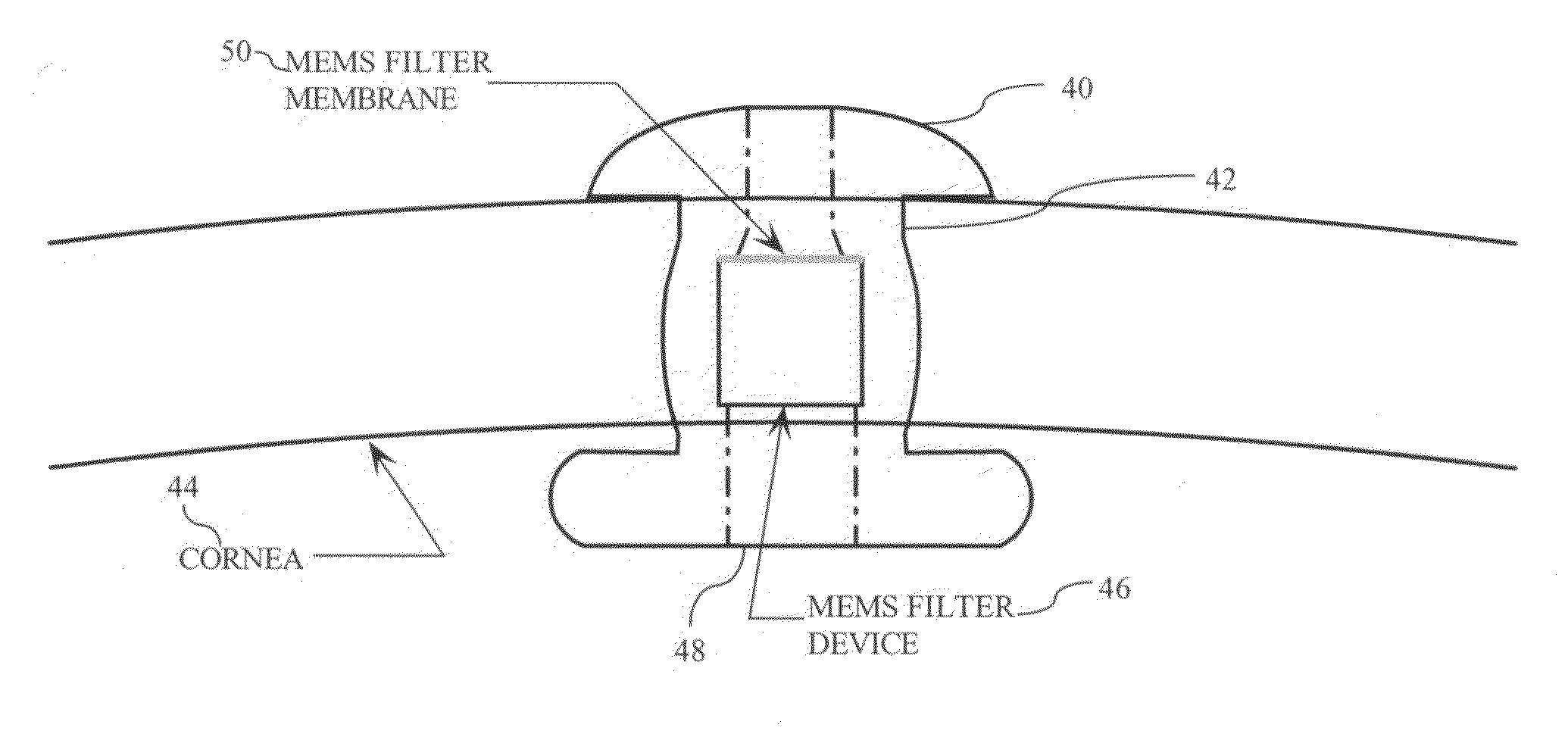
[0039]FIG. 1 illustrates an example of a transcorneal shunt. FIG. 1 shows the shunt 40 inserted through an incision 42 in cornea 44. A Micro-Electro-Mechanical Systems (MEMS) filter device 46, disposed in a central passage 48 of the shunt 40, has a perforated membrane 50 to regulate aqueous humor outflow and limit ingress of microorganisms.

[0040]In some MEMS membrane filter devices, the thin membrane is fabricated to sit on top of a silicon support and is thereby exposed directly to potential damage during handling or assembly. Thus, the mechanical strength of such a membrane is a concern, since it lacks protection for handling or assembly.

[0041]Certain MEMS membrane filter devices may provide a limited flow rate, rendering them ineffective in connection with a glaucom...


PUM


Abstract

A MEMS-fabricated filter device for an ophthalmic shunt and a method for making the same. The filter device may include: a membrane with a plurality of pores, substantially uniformly sized to achieve a therapeutic flow rate while substantially preventing bacterial passage therethrough; a pair of substrates, each bonded to an opposing side of the membrane, and each having an axial inlet opening at a distal end thereof, and a cross-shaped support disposed in one of the substrates, the cross-shaped support supporting the membrane. The filter device may also include: a substrate having a passage therethrough; a membrane, axially recessed from opposing ends of the substrate and having a plurality of pores, substantially uniformly sized to achieve a therapeutic flow rate while substantially preventing bacterial passage therethrough; and a conformal coating covering the membrane. The method may include depositing a membrane layer on a substrate, patterning pores in the membrane layer to define an initial size of the pores, backside etching the substrate to the membrane layer, and conformally coating the membrane layer.

Description

BACKGROUND OF THE INVENTION[0001]1. Field of the Invention[0002]The present invention relates to a filter device for a medical device and manufacturing methods thereof. More particularly, certain implementations of the invention provide for a MEMS-fabricated filter device and / or flow restricting device (and manufacturing methods thereof) for an ophthalmic shunt for implantation through the cornea or sclera of an eye to relieve intraocular pressure in the anterior chamber, and for implantation through the sclera to introduce medications into the posterior chamber. As such, the embodiments of the present invention are useful, for example, in both transcorneal and transscleral applications.[0003]2. Description of the Related Art[0004]Glaucoma, a condition caused by optic nerve cell degeneration, is the second leading cause of preventable blindness in the world today. A major symptom of glaucoma is a high intraocular pressure, or “IOP,” which is caused by the trabecular meshwork failing...

Claims


Application Information

Patent Timeline
Patent Type & Authority Applications(United States)
IPC IPC(8): B01D39/14
CPCA61F9/00781B01D67/0062B01D71/02B01D2325/028B01D2325/48B01D71/0213B01D71/0215
Inventor LIU ZHIXIONG (ERIC)
Owner BECTON DICKINSON & CO
Why Eureka
Try Eureka