

Production from blood of cells of neural lineage
a neural lineage and blood technology, applied in the field of neural lineage blood cell production, can solve the problems of limited neuroscientists' success in developing therapies, inability of the central nervous system (cns) to substantially regenerate,
- Summary
- Abstract
- Description
- Claims
- Application Information
AI Technical Summary
Benefits of technology
Problems solved by technology
Method used
Image

Examples
example 5
Cellular Immunostaining of the NPCP Neural Lineage Markers
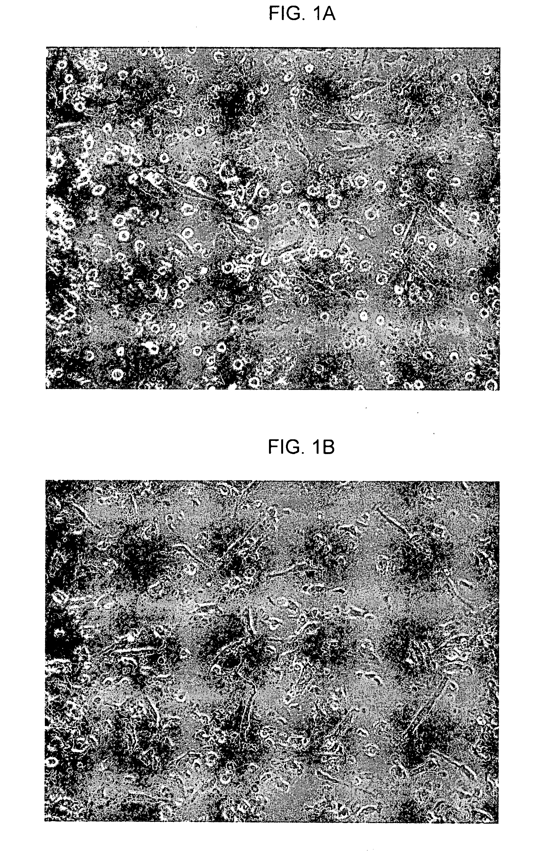
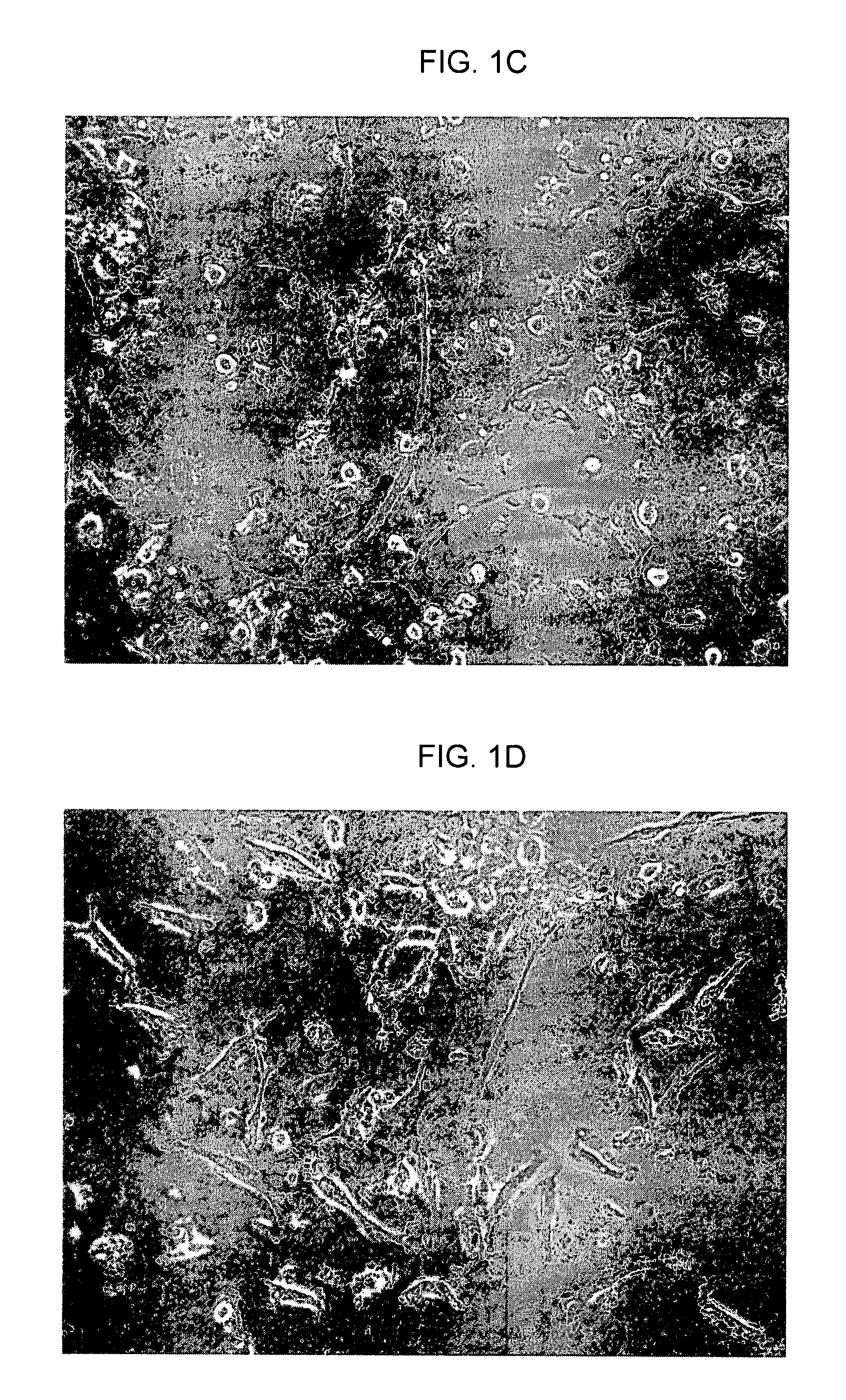
[0315]FIGS. 3A-E are photographs showing immunostaining results for a CCP-derived NPCP, in accordance with an embodiment of the present invention. In the same set of experiments that produced the results shown in FIG. 1, immunostaining of neural lineage markers was performed. Slide-fixed NPCP cells were stained with: FIG. 3A—Anti-Neu-N-Alexa 488; FIG. 3B—Anti-nestin detected by anti-mouse IgG-FITC; FIG. 3C—Anti-tubulin detected by anti-mouse IgG-FITC; FIG. 3D-Anti-04 detected by anti-mouse IgG-Cy3; and FIG. 3E. Anti-GFAP detected by anti-mouse IgG-Cy3. Cells stained with non-specific mouse IgG were detected by anti-mouse IgG-FITC or by anti-mouse IgG-Cy3 used as negative controls.
[0316]Peripheral blood was extracted from nine normal human donors for use in nine respective experiments. The in vitro generation of the NPCP was carried out in accordance with an embodiment of the present invention. Images in FIG. 3 show that NPCP ...
example 6
Flow Cytometry Analysis of the NPCP Neuronal Markers Nestin and β-Tubulin
[0317]FIG. 4 is a graph showing the percentage of cells testing positive (+ / −1 S.E.) for the neural markers Nestin and β-Tubulin, in accordance with an embodiment of the present invention. In the same set of experiments that produced the results shown in FIGS. 1, 2 and 3, cellular immunostaining of the neuronal markers Nestin and β-Tubulin was performed at several time points (days 0, 5, 12, 19 and 26) during the culture period. Staining was performed by applying specific antibodies or normal mouse IgG1 (negative isotype control) detected by PE-conjugated anti-mouse antibodies. The in vitro generation of the NPCP was carried out in accordance with an embodiment of the present invention.
[0318]The figure shows that Nestin-exhibiting cells can be detected earlier during the culture period (starting form day 5) while β-Tubulin levels are elevated on the cells starting from day 12. The percentages of cells exhibitin...
example 7
NPCP Cells Physiologically Respond to Neurotransmitter Stimulation In Vitro
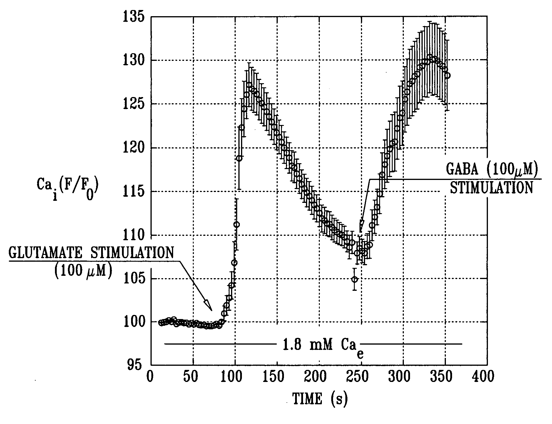
[0319]FIG. 5 is a graph showing typical average calcium levels generated by CCP-derived NPCP cells in response to stimulation by the neurotransmitters glutamate and GABA, in accordance with an embodiment of the present invention. In the same set of experiments that produced the results shown in FIGS. 1-4, NPCP cells' physiological response to neurotransmitters was measured. The in vitro generation of the NPCP was carried out in accordance with an embodiment of the present invention. Ca2+ influx through voltage-gated calcium channels in response to neurotransmitter stimulation with 100 μM glutamate and 100 μM GABA was measured. Harvested cells were loaded with 5 mM Fura-2 acetoxymethyl ester and mounted in a chamber that allowed the superfusion of cells in analytes. Fura-2 was imaged and fluorescent imaging measurements were acquired. The NPCP cells' response to stimulation with the neurotransmitters glutamate...
PUM
| Property | Measurement | Unit |
|---|---|---|
| density | aaaaa | aaaaa |
| density | aaaaa | aaaaa |
| density | aaaaa | aaaaa |
Abstract
Description
Claims
Application Information