Medical image display apparatus, method and program, and recording medium for the program

a technology for medical images and recording media, applied in the field of medical image display apparatus, method and program, and recording media for program, can solve problems such as inability to meet the needs, and achieve the effect of improving image interpretation efficiency and easy image interpretation

Active Publication Date: 2009-01-15
FUJIFILM CORP
View PDF6 Cites 95 Cited by
  • Summary
  • Abstract
  • Description
  • Claims
  • Application Information

AI Technical Summary

Benefits of technology

[0028]According to the present invention, it is possible to automatically display a plurality of slice images including an abnormal shadow region from a series of slice images constituting a three-dimensional medical image in a display mode that is efficient and enables easy image interpretation, thereby enabling contributing to the improvement of the efficiency of image interpretation.

Problems solved by technology

However, the medical image management and display system described in Patent Document 1 has a problem in that it requires a user to provide display mode information to each medical image in advance, which is cumbersome.
Furthermore, in recent years, since numerous slice images are taken at one time by means of a multi-slice X-ray CT apparatus or the like, efficient image interpretation is needed, but the inventions described in Japanese Patent Application Laid-Open No. 2005-334634 and Patent Application Laid-Open No. 2002-143095 cannot meet that need.

Method used


Image

  • Medical image display apparatus, method and program, and recording medium for the program
  • Medical image display apparatus, method and program, and recording medium for the program
  • Medical image display apparatus, method and program, and recording medium for the program

Examples


first embodiment

[0059]FIG. 4 is a flowchart illustrating a first embodiment of a medical image display method according to the present invention, and this processing is executed by starting a medical image display program.

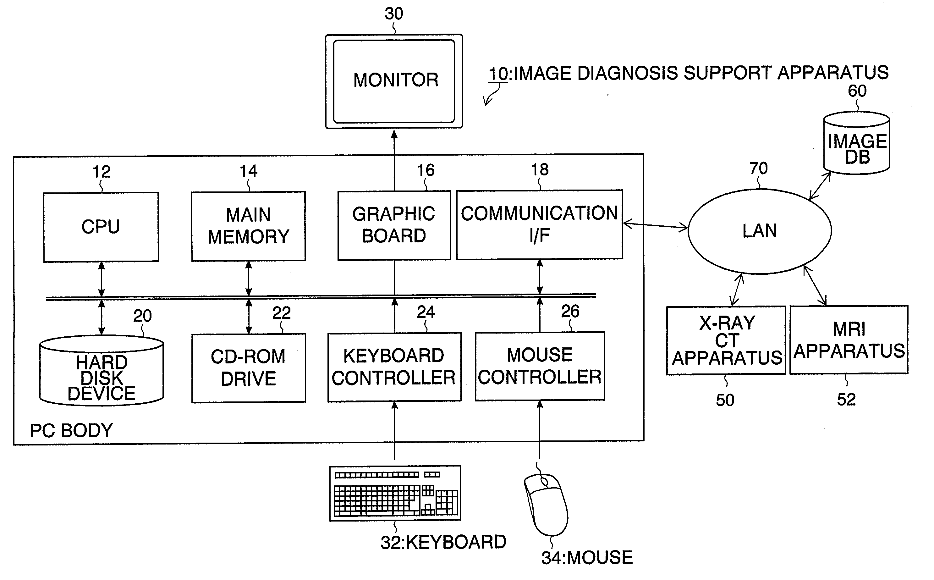
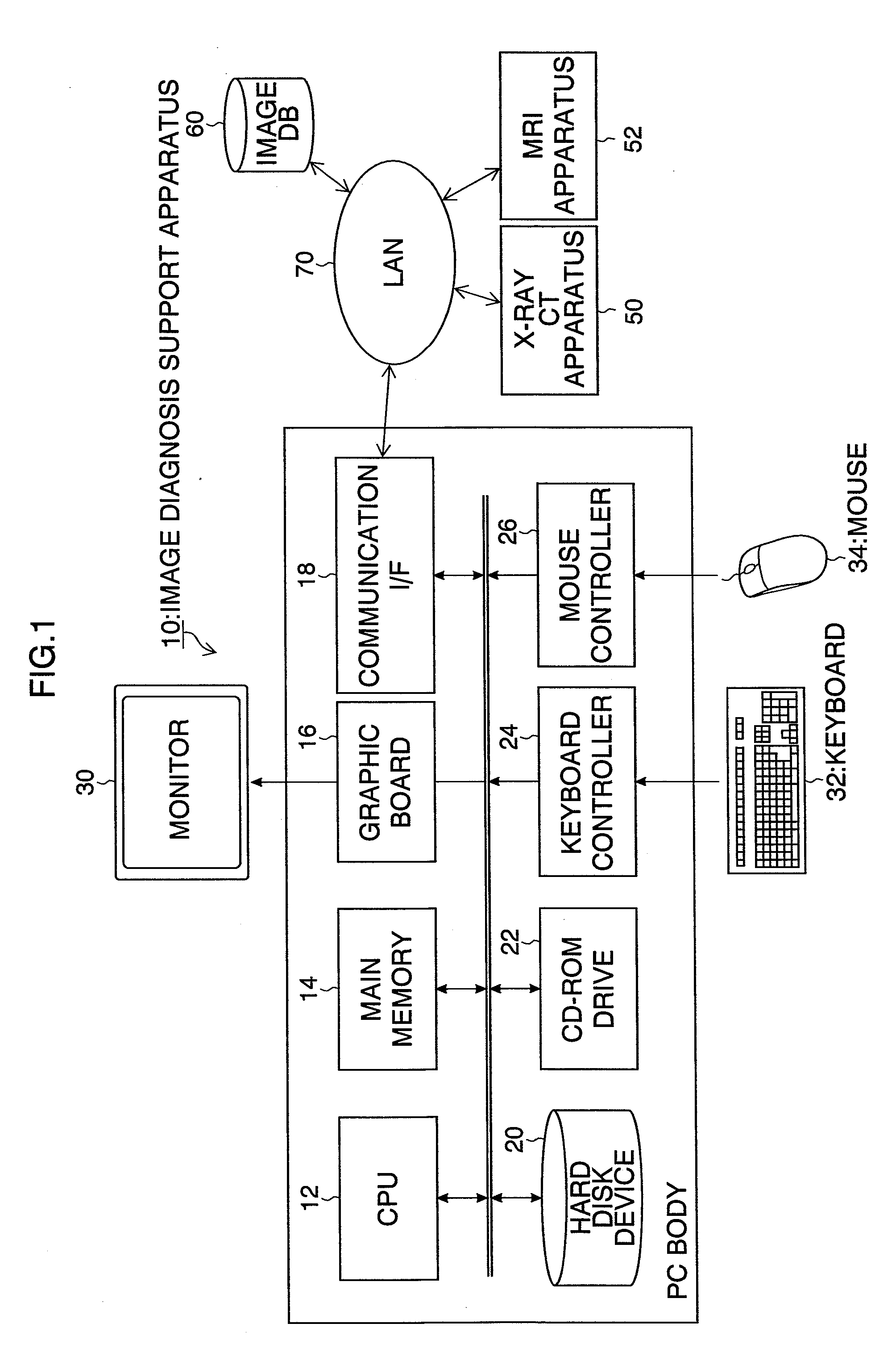
[0060]A diagnostic radiologist operates the keyboard 32 and / or the mouse 34 of the medical image display apparatus 10 to input a patient's name, a date and time of the image-taking, a body part of which the images were taken and a 3D modality type, etc., and acquires a group of slice images (a series of slice images), which is the interpretation target, from the image DB 60 based on this input information (step S10).

[0061]Subsequently, multi-screen display of a plurality of slice images from the acquired series of slice images is provided on the monitor device 30 (step S12). Here, when a 15-split screen in which one screen is split into 3×5 segments is set as a split screen for multi-screen display of the plurality of slice images, 15 slice images, i.e., the first to fifteenth sli...

second embodiment

[0091]FIG. 12 is a flowchart illustrating a second embodiment of a medical image display method according to the present invention. Steps that are common to those in the first embodiment shown in FIG. 4 are provided with the same step numerals, and the detailed description thereof will be omitted.

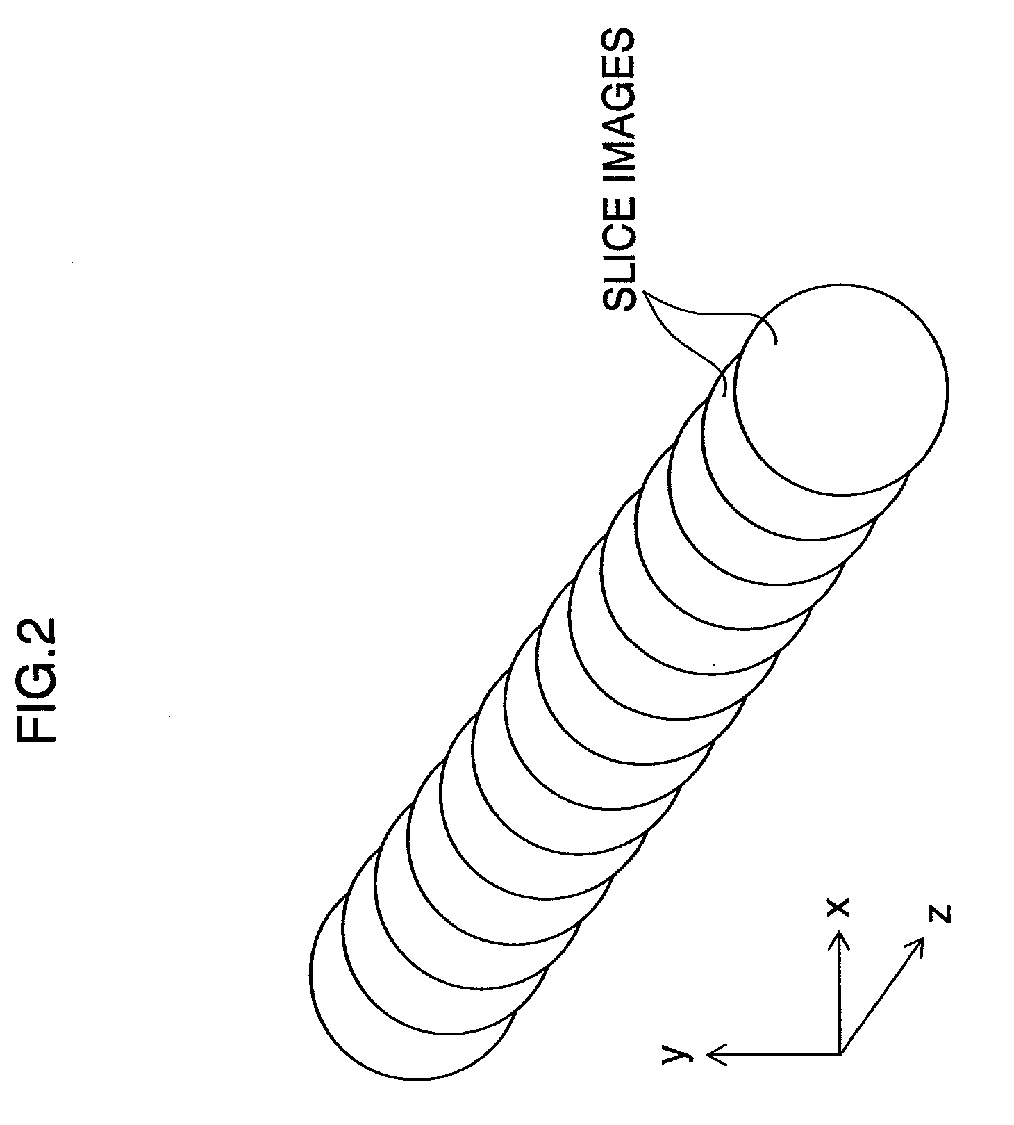
[0092]Upon acquisition of 3D information representing the outer shape of an abnormal shadow region by means of step S14, based on this 3D information, a slice image in the center position of a plurality of slice images including the abnormal shadow region (slice image at the middle of the length in the slicing-proceeding direction (z-axis direction) of the abnormal shadow region) is obtained.

[0093]Then, with reference to the slice image at the middle of the length in the slicing-proceeding direction of the abnormal shadow region, the layout of the plurality of slice images including the abnormal shadow region on the split screen is determined, and a display screen for providing multi-screen...

third embodiment

[0096]FIG. 13 is a flowchart illustrating a third embodiment of a medical image display method according to the present invention. Steps that are common to those in the first embodiment shown in FIG. 4 are provided the same step numerals, and the detailed description thereof will be omitted.

[0097]Upon acquisition of 3D information representing the outer shape of an abnormal shadow region by means of step S14, the first slice image in a plurality of slice images including the abnormal shadow region is obtained based on this 3D information. For example, a slice image with a smallest positional value in the slicing-proceeding direction (z coordinate) in the plurality of slice images including the abnormal shadow region is obtained, or since the slice images are provided with slice numbers for respective slicing positions in the z-axis direction, a slice image provided with the smallest slice number in the plurality of slice images including the abnormal shadow region abnormal shadow re...


PUM


Abstract

According to the medical image display apparatus of an aspect of the present invention, the display mode to provide multi-screen display of the plurality of slice images including the abnormal shadow region is determined based on the result of acquisition of the abnormal shadow region (three-dimensional information for the abnormal shadow region), which eliminates the necessity for users (doctors) to perform an operation to set a display mode, and enables display of the plurality of slice images including the abnormal shadow region efficiently in a display mode enabling easy image interpretation.

Description

BACKGROUND OF THE INVENTION[0001]1. Field of the Invention[0002]The present invention relates to a medical image display apparatus, method and program, and recording medium for the program. More specifically, the present invention relates to a technique for displaying on a multi-screen a series of slice images taken by means of an X-ray CT apparatus or an MRI apparatus, etc.[0003]2. Description of the Related Art[0004]Conventionally, a medical image management and display system configured so that an image file containing medical images is provided with display mode information having information for a split screen and medical image frame positions, and when displaying the medical images contained in this image file on a viewer, the screen of the viewer is split according to the split screen information included in the display mode information, and the medical images are displayed at the corresponding positions in the split screen according to the frame position information included...

Claims


Application Information

Patent Timeline
Patent Type & Authority Applications(United States)
IPC IPC(8): H05G1/64
CPCA61B5/055A61B6/032A61B6/463A61B6/466A61B6/566G06T2207/30096G06F19/3406G06T7/0012G06T2200/24G06T2207/10072G06F19/321G16H30/20G16H30/40G16H40/63G16H50/20
Inventor LI, YUANZHONG
Owner FUJIFILM CORP
Why Eureka
Try Eureka