
Airway intubation device

a technology of airway and intubation, which is applied in the field of medical devices, can solve the problems of patient's death, failure to achieve 90% success rate, and failure to achieve oxygenation, so as to avoid tissue damage during intubation and improve the illumination of the intubation operation.

Active Publication Date: 2009-02-05
AMBU AS
View PDF26 Cites 74 Cited by
  • Summary
  • Abstract
  • Description
  • Claims
  • Application Information

AI Technical Summary

Benefits of technology

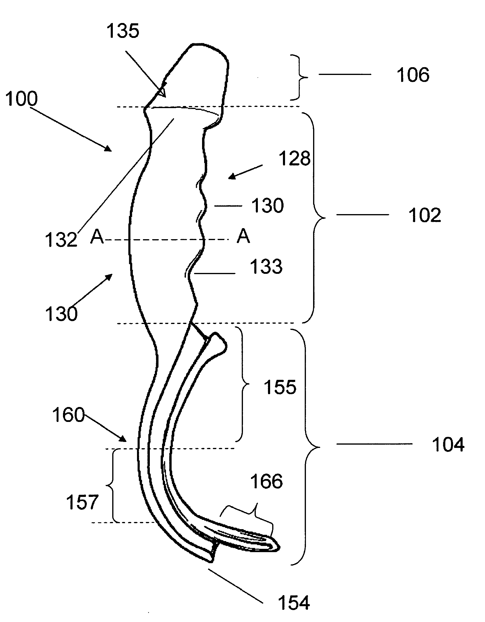
The invention is an airway intubation device that allows for easy and safe insertion of a tube into a patient's trachea during medical procedures. The device is made from a single rigid body and includes a hand piece with a comfortable grip, a cap piece, and an insertion piece with a curved design for better control. The insertion piece has a wide entry portal and a narrower slot to keep the tube in place. The device also includes an illumination system for better visibility during the procedure and an image viewing system for real-time guidance. The video display screen includes an anatomically correct aiming reticle for accurate placement of the tube. The device is made from a biocompatible material and can be molded from stainless steel or other materials.

Problems solved by technology

Unrecognized failure to correctly place an endotracheal tube into the trachea can result in a failure of oxygenation, and the patient's death.
A real challenge remains in that it takes 50 or more intubations to achieve a 90% success rate.
Using these devices, visualization of the vocal cords is often made easier but actual delivery of the breathing tube (endotracheal tube, ETT) between the cords into the windpipe (trachea) of the patient often remains difficult.
When using these devices, not infrequently, during navigation of the ETT to and through the vocal cords, forward advancement of the ETT can be impeded by ‘hang-up’ on the right vocal cord.
In addition, sporadic cases of trauma to the soft palate have been reported.
However, while the delivery channel was an attempt to deal with the challenges of tube placement, success was often impaired because: (a) the rounded (tubular) blade was too narrow to control the tongue and soft tissues well, so that one could often ‘get lost’ and not see the target vocal cords, and (b) the tube emerged from the delivery channel heading in an angle that often resulted in the tube being directed incorrectly into the esophagus and not the trachea.

Method used


Image

  • Airway intubation device
  • Airway intubation device
  • Airway intubation device

Examples


Embodiment Construction

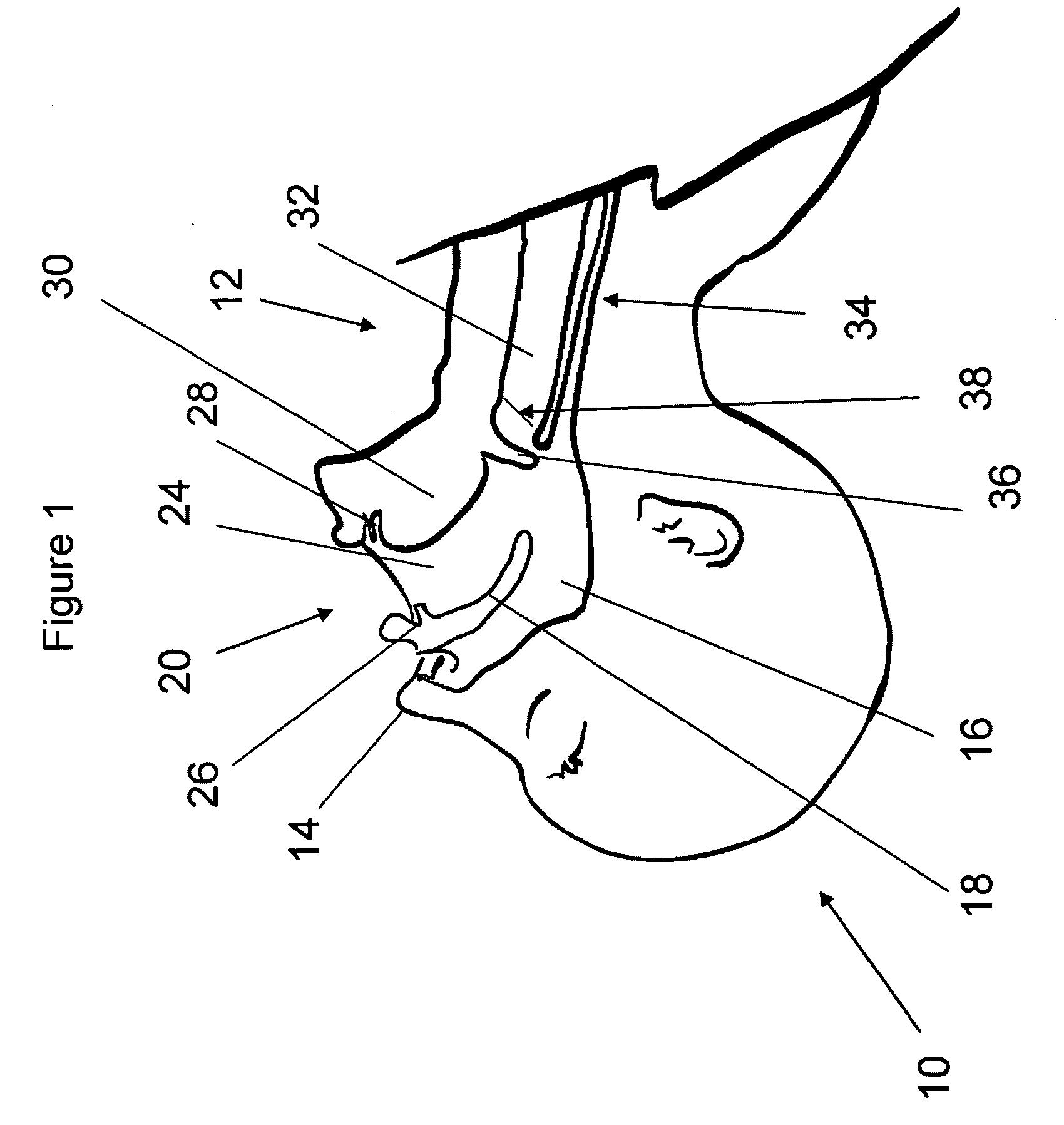
[0037]The process and principals of endotracheal intubation are well known. Referring to FIG. 1, there is shown a cross-section of a human head (10) and neck (12) to illustrate the anatomy relevant to intubation. There is shown the nose (14) and nasal passage (16). The palate (18) separates the mouth (20) and oral cavity (24) from the nasal passages. The upper (26) and lower (28) teeth and tongue (30) are also shown. During breathing, air travels down the windpipe or trachea (32) into the lungs. Food and water travel down the esophagus (34) to the stomach. To avoid choking, the epiglottis (36) closes the trachea when swallowing and opens again to permit breathing. The vocal cords (38) are located below the epiglottis and mark the entry point into the trachea. All of these airway soft-tissues are vulnerable to damage during intubation. The major challenge in performing endotracheal intubation relates to the need to see around the corner created in part by the tongue and epiglottis. T...


PUM


Abstract

An airway intubation device having a one-piece molded body of biocompatible material with an ergonomically designed hand piece for left hand manipulation. The body includes a cap piece and linear-curved-linear insertion piece having a channel in one side thereof for insertion of an endotracheal tube. The channel has a wide entry portal disposed ahead of the hand piece so that a clinician may load the channel with a tube while the insertion piece is in the mouth of the patient without having to view the entry portal. The device includes illumination means and viewing means comprising an eye piece or a video display unit mounted to the cap piece so that a clinician can view the insertion of the tube between the vocal cords of a patient without contact.

Description

RELATED APPLICATIONS[0001]This application claims the benefit of provisional patent application No. 60953992 filed in the United States Patent and Trademark Office on Aug. 4, 2007.BACKGROUND OF THE INVENTION[0002]1. Field of the Invention[0003]This invention relates to medical devices and more specifically to an improved airway intubation device.[0004]2. Background of the Invention—the Problem to be Solved[0005]In both acute-care medicine and as part of delivering a general anesthetic for an operative procedure it is often necessary to insert a breathing tube into the airway of a living organism, most often a human, but in some cases this can also apply to pets, livestock and other animals. This procedure is called endotracheal intubation. A variety of tools exist for performing this procedure, which is usually performed by highly trained professionals in a hospital, or pre-hospital setting. Most intubations are performed with direct laryngoscopy, using a device called a direct lary...

Claims


Application Information

Patent Timeline
Patent Type & Authority Applications(United States)
IPC IPC(8): A61M16/00
CPCA61M16/0488A61B1/00032A61B1/00052A61M16/0495A61B1/0684A61B1/2673A61M2205/32A61B1/04
Inventor LAW, JOHN ADAMGROSS, MICHAELKOVACS, GEORGE JAMESCARKNER, STEVE
Owner AMBU AS
Learn More