

Protein Extraction buffer, a kit comprising it and method of its use
a protein extraction and protein technology, applied in the field of proteomics, can solve the problems of insufficient preservation of antibody epitopes in ffpe tissue sections, especially difficult protein extraction of ffpe tissue sections, and inability to achieve protein extraction buffers to achieve the preservation of antibody epitopes. , to achieve the effect of preserving protein epitopes
- Summary
- Abstract
- Description
- Claims
- Application Information
AI Technical Summary
Benefits of technology
Problems solved by technology
Method used
Image

Examples
example 1
Deparaffinization and Protein Extraction from FFPE Tissue Sections
[0042]FFPE slides are obtained from any source, such as a pathology lab. Section from FFPE slides are up to about 10 μm thick. Up to 2 sections, each with a thickness of about 10 μm and area of up to about 100 mm2, are combined in one preparation. Smaller sections are also combined for one preparation. The yield of extracted protein depends on the amount and the nature of the starting material and may vary depending on cell type.
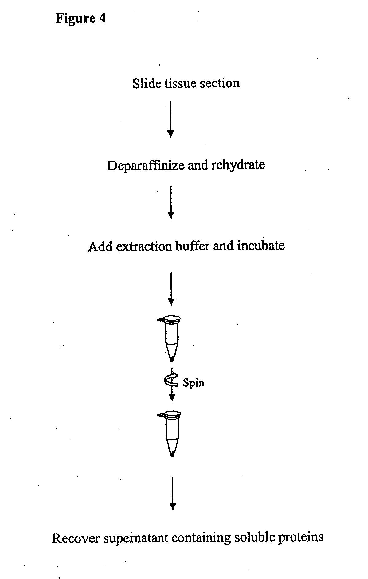
A. Deparaffinization
[0043]Before deparaffinization, slides are kept at room temperature for 60 min. or heated at 60° C. for 20 min. in a horizontal position. The slide is immersed in xylene for 10 min. This step is repeated once in new xylene solution for 10 min. The slide is then immersed in 100% ethanol for 5 min.; followed by another immersion in 85% ethanol for 1 min.; followed by yet another immersion in 70% ethanol for 1 min. The slide is then immersed in high purity water for 1 min. Exc...
example 2
Comparison of Proteins Obtained From Breast Tumor Samples Using Different Extraction Buffers
A. Deparaffinization of Tissue Sections
[0053]For each extraction condition, 2 slides of the same breast tumor patient (HuCAT295, USBiomax, Rockville Md.) were used. To deparaffinize FFPE tissue sections, slides were immersed in xylene for 5 min.; in fresh xylene for 5 min.; in 100% ethanol for 5 min.; in 85% ethanol for 1 min.; in 70% ethanol for 1 min.; and in high purity water for 1 min. Excess water was removed and tissue sections were transferred to a clean Eppendorf tube using a razor blade by pooling 2 tissue sections together.
B. Protein Extraction
[0054]35 μl ABP extraction buffer (of the invention) was added to the tissue section. The tissue was broken using a plastic pestle and heated at 100° C. for 20 min. After a quick spin down, the tissue was broken again with the plastic pestle and incubated at 60° C. for 2 hours. The sample was then centrifuged at 15,000 g for 10 min. at 4° C. a...
example 3
Comparison of Extraction Buffers for FFPE
A. Extraction Buffer of the Invention
[0059]Two 10 μm tissue section slides were taken from the same patient. The slides were immersed in xylene for 5 min. and immersed again in fresh xylene solution for 5 min. The slides were then immersed in 100% ethanol for 5 min. This step was repeated. The slides were then immersed in 85% ethanol for 1 min., followed by immersion in 70% ethanol for 1 min. The slides were then immersed in water for 1 min. Excess water was removed from the slides and the sections are transferred to a clean Eppendorf tube using a razor blade.
[0060]35 μl Extraction Buffer was added to one Eppendorf tube containing the tissue section. The sample tube was heated at 100° C. for 20 min. The sample tube was centrifuged to spin down the liquid. The sample tube was then incubated at 60° C. for 2 hours, and vortexed every 30 min. during the incubation. The sample tube was then centrifuged for 10 min. at 15,000×g at 4° C. The soluble ...
PUM
| Property | Measurement | Unit |
|---|---|---|
| Temperature | aaaaa | aaaaa |
| Temperature | aaaaa | aaaaa |
| Molar density | aaaaa | aaaaa |
Abstract
Description
Claims
Application Information