Registration method with three-dimensional representation of a vascular tree as a function of blood flow

Active Publication Date: 2009-03-19
SIEMENS HEALTHCARE GMBH
View PDF1 Cites 19 Cited by
  • Summary
  • Abstract
  • Description
  • Claims
  • Application Information

AI Technical Summary

Benefits of technology

[0012]The object of the present invention is to create a method for determining a set of imaging parameters, which allows high-quality registration. Registration here should be possible, regardless of whether a biplane x-ray system or a monoplane x-ray system is used, whether or not an individual DSA image is available with a high fill level, etc. It should also not be necessary to determine a summation image based on the DSA sequence.

Problems solved by technology

However both modalities also have specific disadvantages.
Registration is often problematic, since a vascular tree that is permanently filled in its entirety with contrast agent is used for the reconstruction of the three-dimensional volume data record.
This significantly impedes registration, as non-identical states have to be linked to one another.
Methods for compensating for such motion are known but compensation is difficult and only possible to a limited degree.
However this procedure involves a significant computation outlay, as the artificial projection has to be determined as well as the determination of the summation image.

Method used


Image

  • Registration method with three-dimensional representation of a vascular tree as a function of blood flow
  • Registration method with three-dimensional representation of a vascular tree as a function of blood flow
  • Registration method with three-dimensional representation of a vascular tree as a function of blood flow

Examples


Embodiment Construction

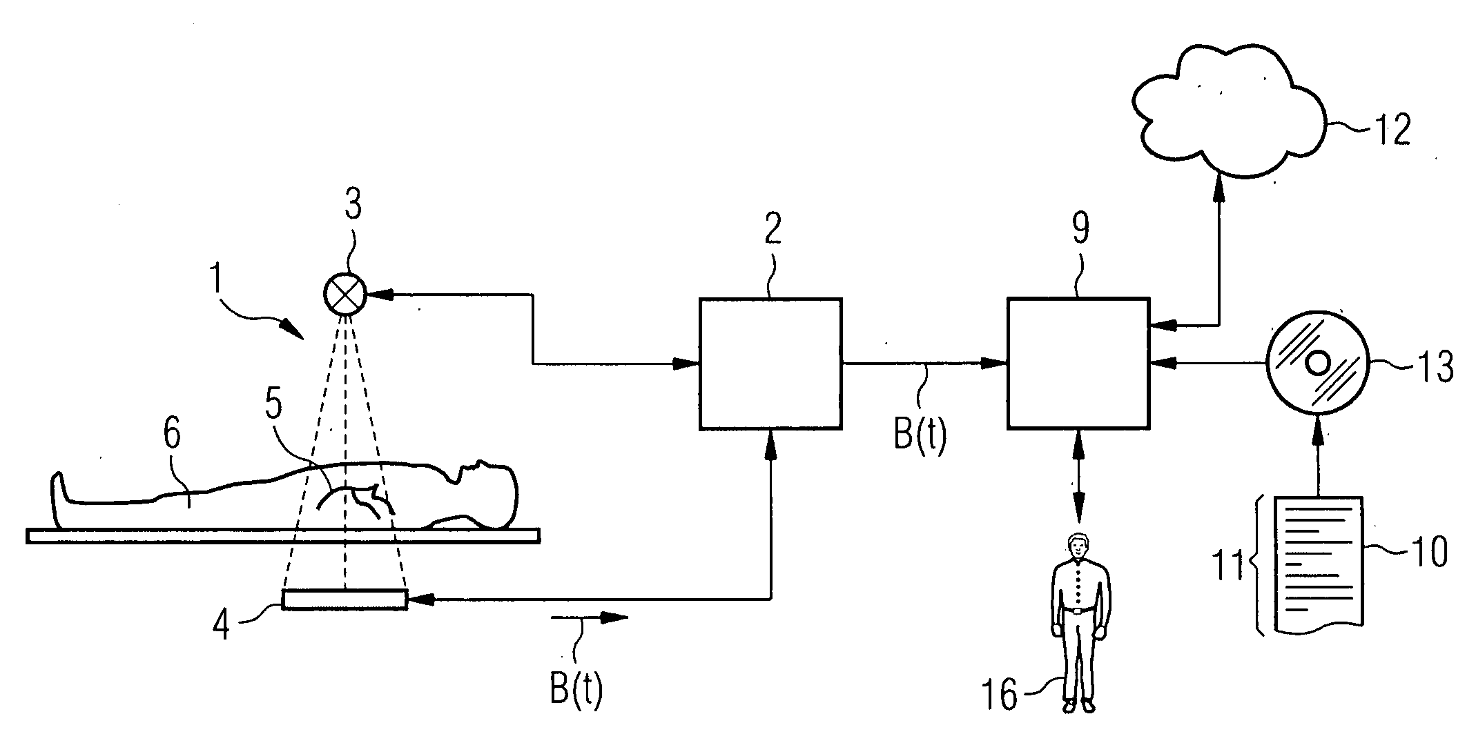
[0036]According to FIG. 1 an x-ray system has an acquisition facility 1 and a control facility 2. The acquisition facility 1 has an x-ray source 3 and an x-ray detector 4. The acquisition facility 1—after corresponding activation by the control facility 2—acquires a sequence of two-dimensional projection images B. Each of the projection images B is acquired here at a respective acquisition time t. The sequence of projection images B shows the flow of a contrast agent through an actually present (and of course three-dimensional) vascular tree 5. The vascular tree 5 is only shown schematically in FIG. 1. It can be present for example in the brain or in another part of the body of a patient 6. The sequence of projection images B is received by the control facility 2 and buffered there.

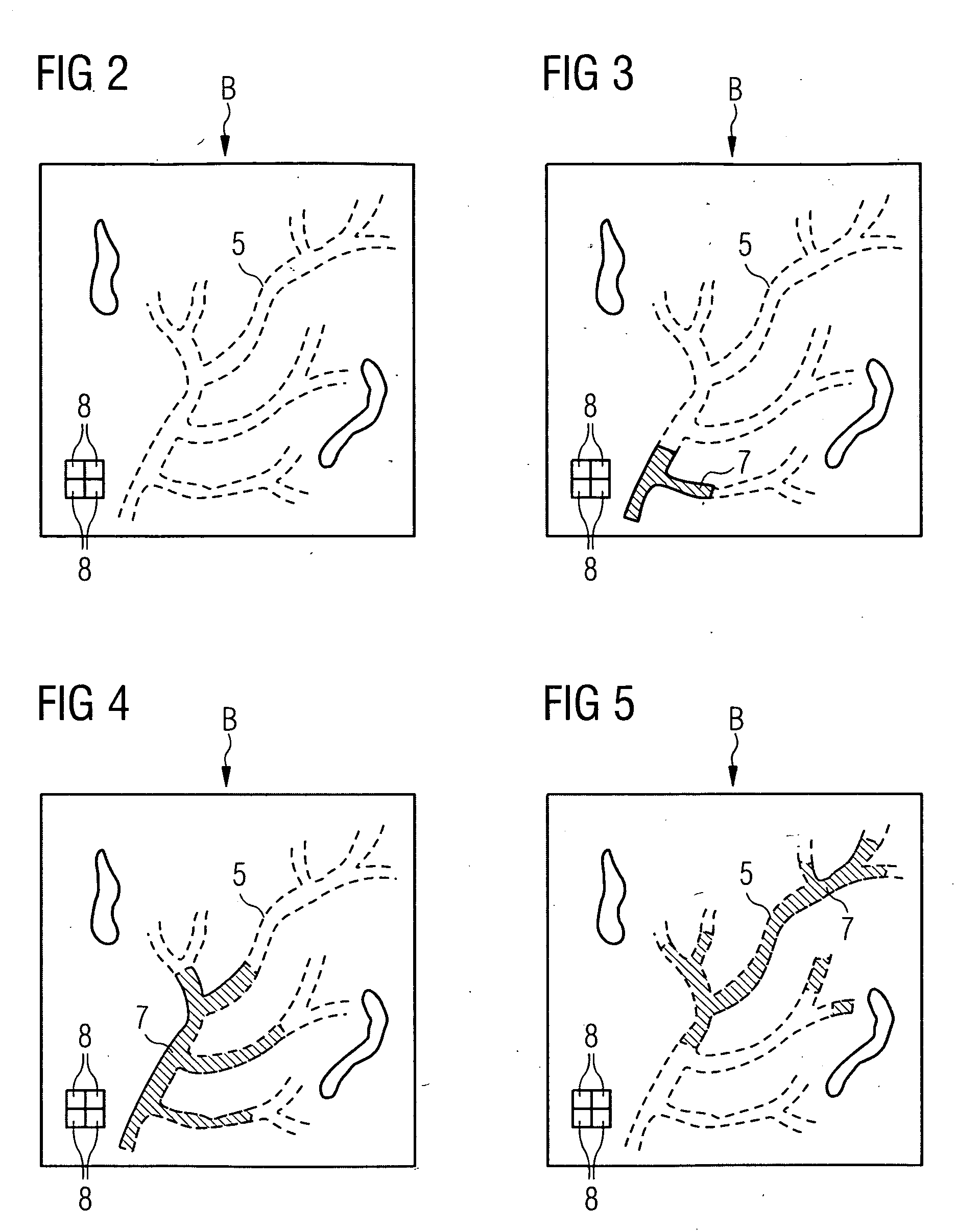
[0037]FIGS. 2 to 5 shows some of the projection images B in the sequence in a highly schematic manner and purely by way of example.

[0038]FIG. 2 shows the first projection image B of the sequence. During a...


PUM


Abstract

A three-dimensional volume data record contains a vascular tree. A two-dimensional projection image is an image of an actual fill state, to which the vascular tree is filled with a contrast agent at an acquisition time. The volume data record and the projection image are registered in relation to one another by means of the set of imaging parameters. A computation facility determines an expected target fill state, which describes which parts of the vascular tree should be filled with contrast agent at acquisition time in the three-dimensional volume data record. The computation facility determines the set of imaging parameters based on the target fill state and the projection image.

Description

CROSS REFERENCE TO RELATED APPLICATIONS[0001]This application claims priority of German application No. 10 2007 044 406.2 filed Sep. 18, 2007, which is incorporated by reference herein in its entirety.FIELD OF THE INVENTION[0002]The present invention relates to a registration method with three-dimensional representation of a vascular tree as a function of blood flow.BACKGROUND OF THE INVENTION[0003]The imaging methods generally used for vascular diagnosis include both two-dimensional digital subtraction angiography (DSA) and three-dimensional rotational angiography. Both modalities have specific advantages in respect of diagnostic options. However both modalities also have specific disadvantages. They cannot simply be interchanged with one another.[0004]Certain diagnosis processes require both temporal and structural information about blood flow. In such instances both technologies have to be deployed. However in order for the treating physician to be able to investigate certain fac...

Claims


Application Information

Patent Timeline
IPC IPC(8): G06K9/00
CPCA61B19/52A61B2019/5291G06T7/0028G06T2207/30104G06T2207/10072G06T2207/10116G06T7/0038A61B90/36A61B2090/365G06T7/33G06T7/38
Inventor DEINZER, FRANKPLATZER, ESTHER-SABRINA
Owner SIEMENS HEALTHCARE GMBH
Why Eureka
Try Eureka