
Method for calibrating nuclear medicine diagnosis apparatus

a radiation detection and calibration method technology, applied in the field of radiation detection apparatus calibration, can solve the problems of poor calibration, ring-shaped radiation source having a hollow portion, and the risk of -ray scattering by an aqueous solution inside the cylindrical phantom, so as to improve the quality of tomographic image and increase the accuracy of detection efficiency calibration

Inactive Publication Date: 2009-04-02
HITACHI LTD
View PDF4 Cites 7 Cited by
  • Summary
  • Abstract
  • Description
  • Claims
  • Application Information

AI Technical Summary

Benefits of technology

[0009]According to the present invention, the following effect can be obtained.
[0010]A cylindrical phantom without scattered radiation is achieved in an emulating manner, so that the accuracy on detection efficiency calibration will increase and the quality of a tomographic image will improve.

Problems solved by technology

On the other hand, there is a risk that a γ-ray is scattered by an aqueous solution itself inside the cylindrical phantom.
Although there is a method for correcting the scattered radiation, the method has a disadvantage that the correction results in poor calibration.
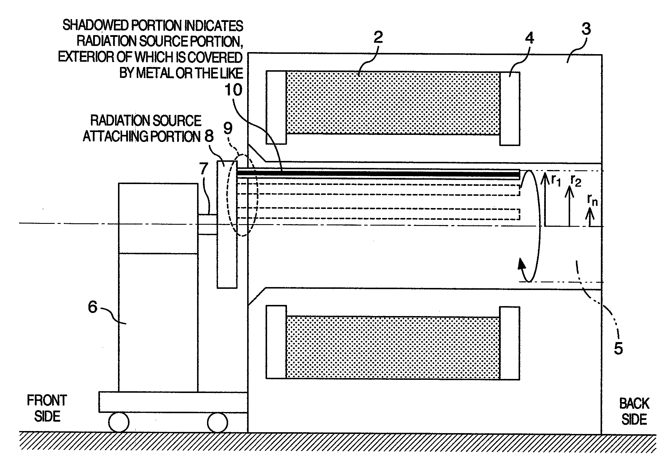
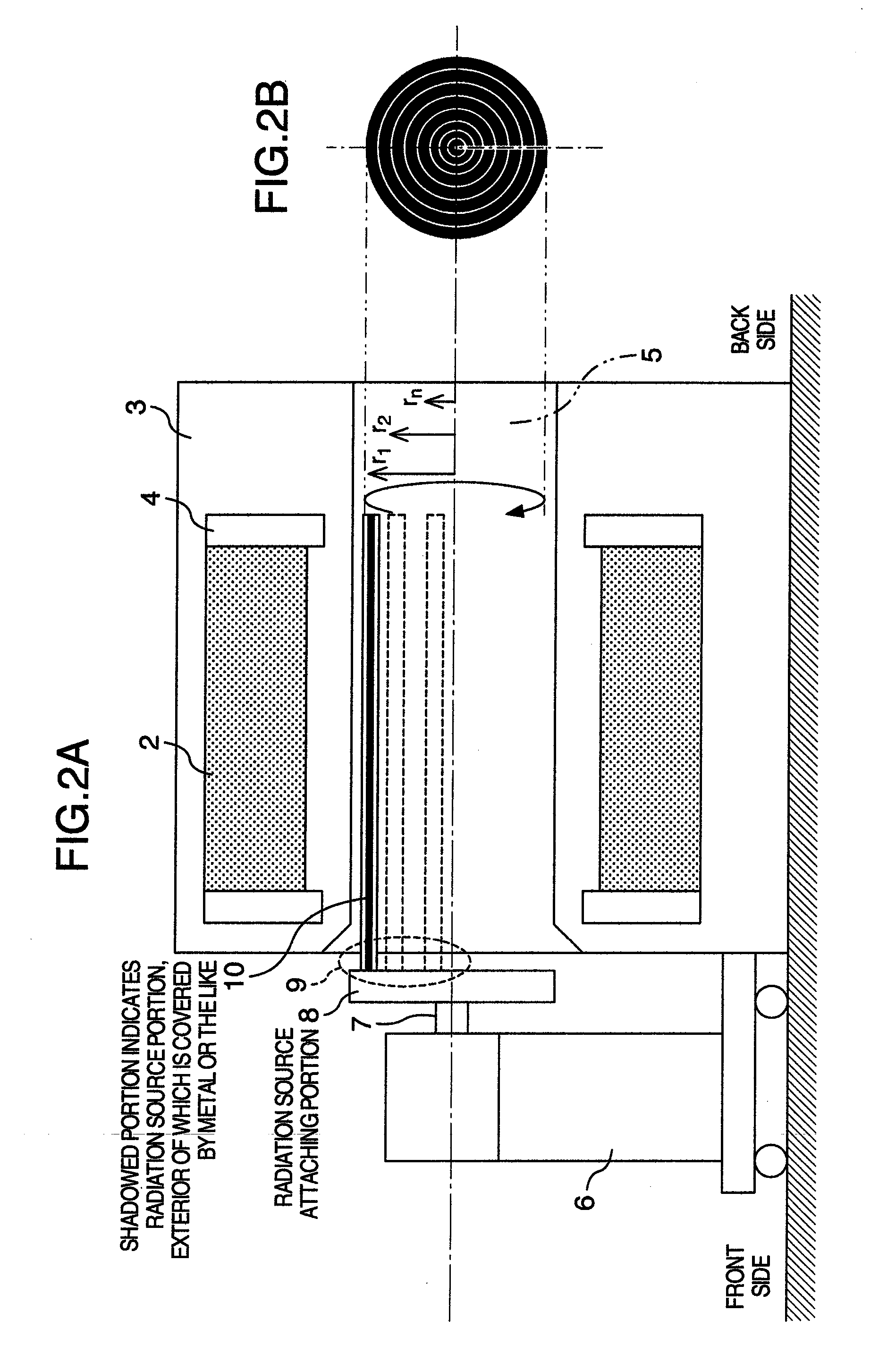
However, this rod-shaped radiation source only rotates on an orbit of a certain radius and thus results in a ring-shaped radiation source having a hollow portion thereinside.
Accordingly, the calibration work will not complete unless two radiation sources are prepared, and this is a very time consuming work.

Method used


Image

  • Method for calibrating nuclear medicine diagnosis apparatus
  • Method for calibrating nuclear medicine diagnosis apparatus
  • Method for calibrating nuclear medicine diagnosis apparatus

Examples


embodiment 1

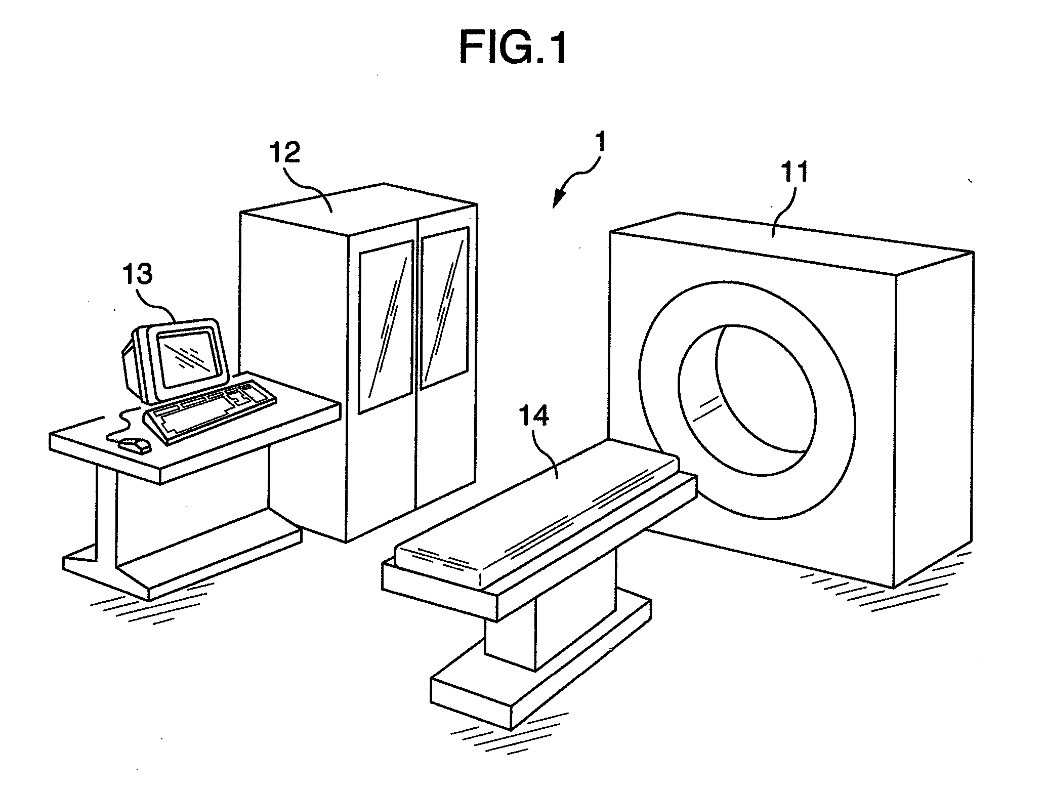
[0024]Any inspection technique using radiation is a technique, in which the physical quantity of a subject to be inspected is measured as an integrated value in the radiation traveling direction, and its integrated value is back projected to calculate the physical quantity of each voxel inside the subject and create an image. In this technique, a large amount of data needs to be processed, and along with the rapid development of computer technologies in recent years, high speed and highly detailed images have come to be provided.

[0025]The X-ray CT technique is a technique, in which the intensity of an X-ray passing through a subject is measured, and from the X-ray transmission coefficient inside the body the morphological information on the subject is imaged. X-rays are emitted from an X-ray source to a subject, whereby the intensity of an X-ray passing through the body is measured with detecting elements disposed opposite to the subject, thereby measuring a distribution of integrat...

embodiment 2

[0050]The rod-shaped radiation source and point radiation source described in Embodiment 1 may be in the form of liquid like a 18F solution instead of in the form of solid. In a rod-shaped radiation source in the form of solid, such as a conventional 68Ge—68Ga, for convenience of the manufacture, radioactivity is likely to fluctuate (e.g., approximately ±10%) in the length direction of the rod, possibly resulting in poor calibration. The use of a liquid radiation source makes the radioactivity spatially uniform and secures more accurate calibration.

[0051]In the calibration method of a nuclear medicine diagnosis apparatus of this embodiment, a liquid radiation source is used as the radiation source, and while rotating the liquid radiation source at a plurality of different turning-radius orbits inside the tunnel of a gantry that collects radiation detection data, the radiation detection data is collected to calibrate the apparatus. Accordingly, other than the advantages of (1) and (2...


PUM


Abstract

A nuclear medicine diagnosis apparatus, which rotates a rod-shaped radiation source or a point radiation source while changing the radius thereof and carries out a calibration work, is provided to resolve the following problems. If detector calibration is carried out using a cylindrical phantom whose interior is filled with a 18F solution, for example, there is a risk that a γ-ray is scattered by the aqueous solution itself inside the cylindrical phantom. Although there is a method for correcting the scattered radiation, the method has a disadvantage that the correction results in poor calibration. Moreover, if a rod-shaped radiation source is rotated inside a tunnel, there is an extremely low risk of causing the above-described scattering because it is rod-shaped. However, this rod-shaped radiation source only rotates on an orbit of a certain radius, resulting in a ring-shaped radiation source having a hollow portion thereinside.

Description

BACKGROUND OF THE INVENTION[0001]The present invention relates to a method for calibrating radiation detection apparatus using radiation.[0002]With an inspection technique using radiation, the interior of a subject can be inspected in a non-destructive manner. In particular, radiographic inspection techniques for a human body include an X-ray CT, a PET, a single photon emission type CT (hereinafter, referred to as “SPECT”: Single Photon Emission Computed Tomography), and the like. In operating the radiographic inspection apparatus, calibrating the apparatus in advance is an essential process in securing the image quality performance of the apparatus. If the calibration has not been carried out sufficiently, a tomographic image may degrade significantly.[0003]As a technique for performing calibration, JP-A-2002-71813 describes a PET including a calibration radiation source P, wherein a rod-shaped radiation source is disposed in a rotating ceptor and revolves in the cylindrical space ...

Claims


Application Information

Patent Timeline
IPC IPC(8): G12B13/00
CPCG01T1/1648
Inventor YANAGITA, NORIHITOINOUE, SHINICHITSUCHIYA, KATSUTOSHI
Owner HITACHI LTD
Learn More