Detection of circulating tumor cells in peripheral blood with an automated scanning fluorescence microscope

Inactive Publication Date: 2009-05-14
IKONISYS INC
View PDF12 Cites 12 Cited by
  • Summary
  • Abstract
  • Description
  • Claims
  • Application Information

AI Technical Summary

Problems solved by technology

The potential interest in the detection of CTC in peripheral blood was first suggested over a century ago (4) but because of their very low numbers CTC cannot be detected by conventional methods.
However, since cell integrity is destroyed during RNA extraction, this approach precludes the analysis of cell morphology and phenotype and so, in particular, may not be able to distinguish material shed directly from normal tissue as opposed to from tumors nor does it allow the detection of several associated changes in the same cell.
RT-PCR tests thus do not enable counting of CTC and can, in general, only give a

Method used


Image

  • Detection of circulating tumor cells in peripheral blood with an automated scanning fluorescence microscope
  • Detection of circulating tumor cells in peripheral blood with an automated scanning fluorescence microscope
  • Detection of circulating tumor cells in peripheral blood with an automated scanning fluorescence microscope

Examples


Embodiment Construction

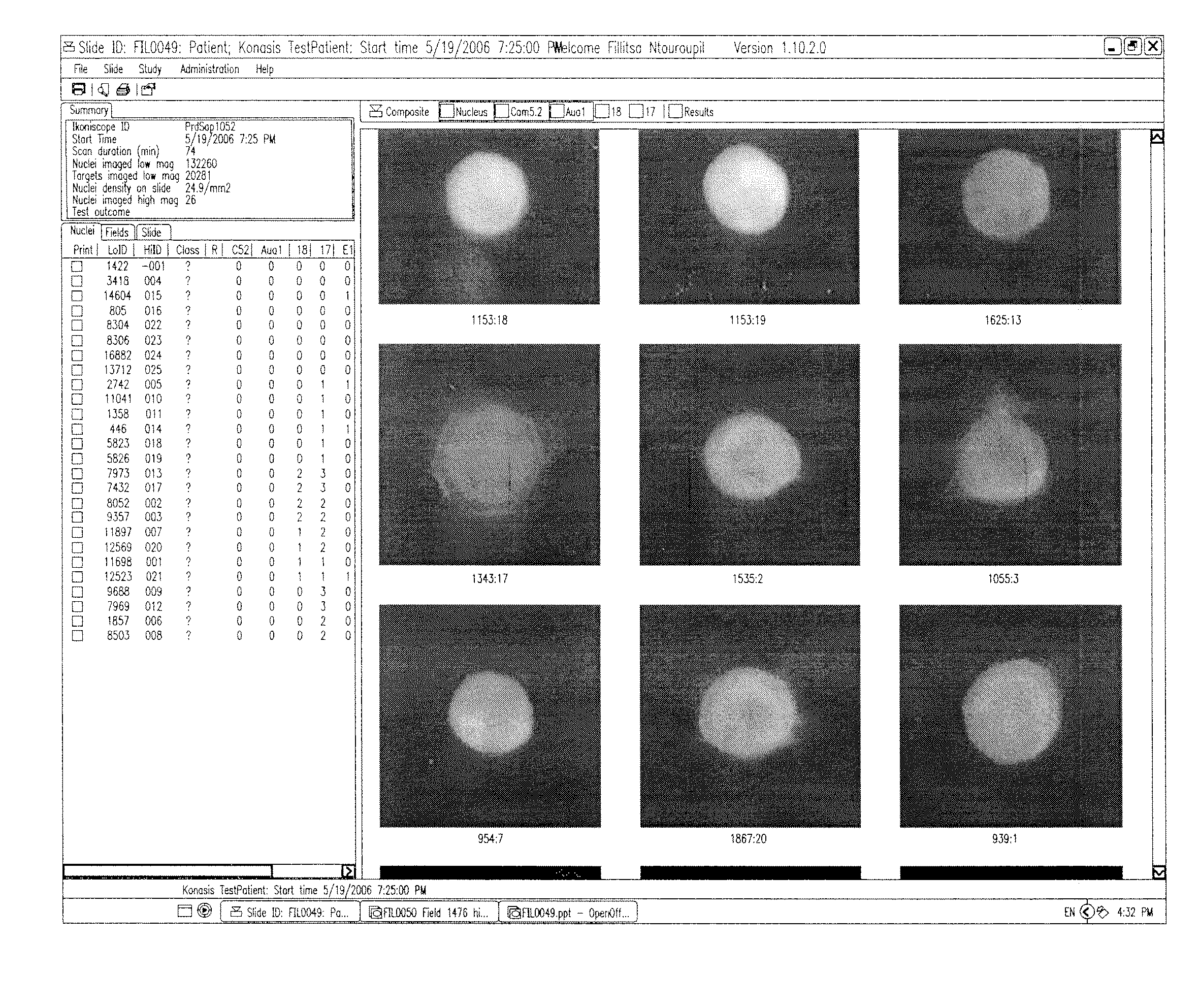
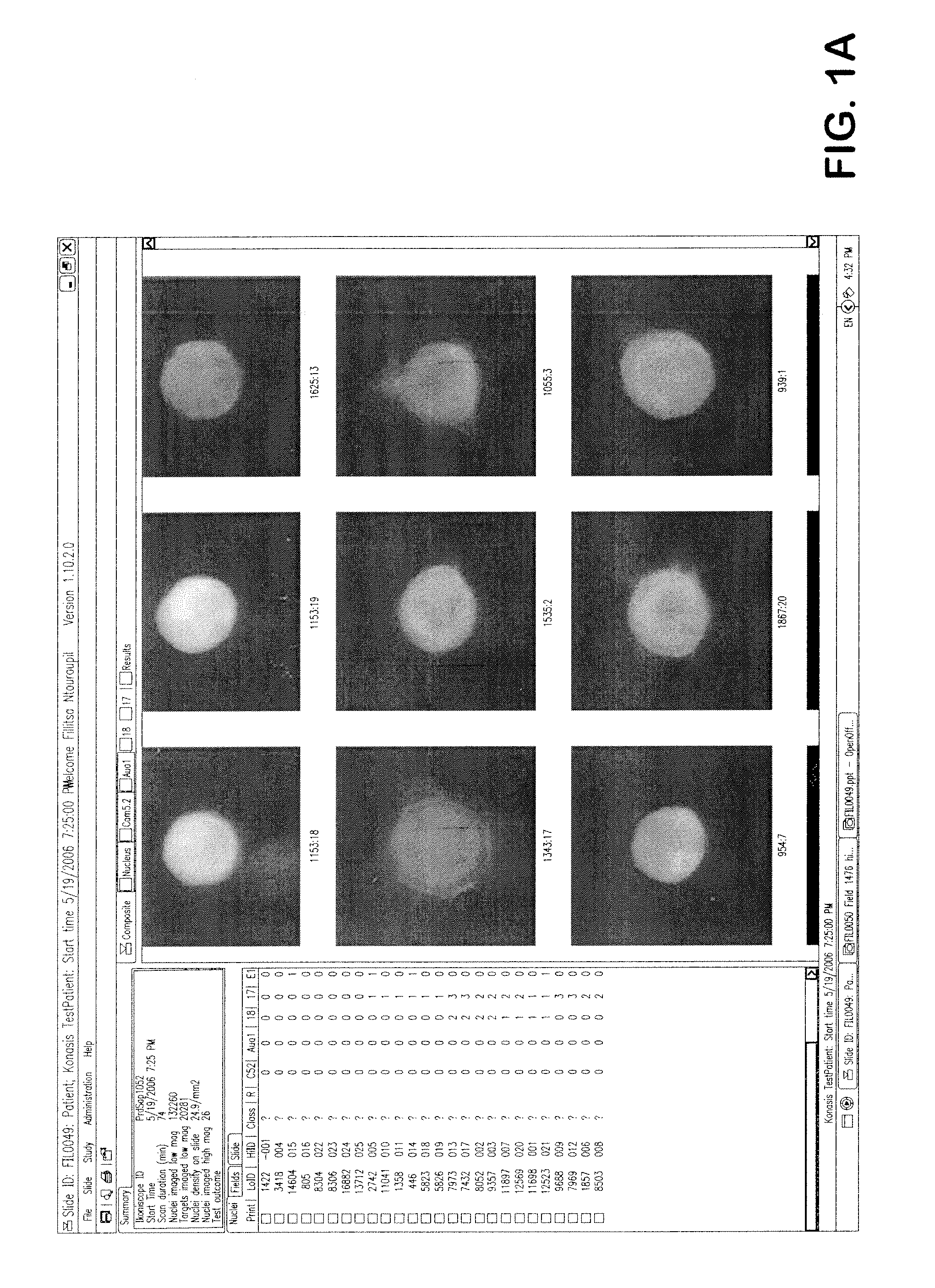
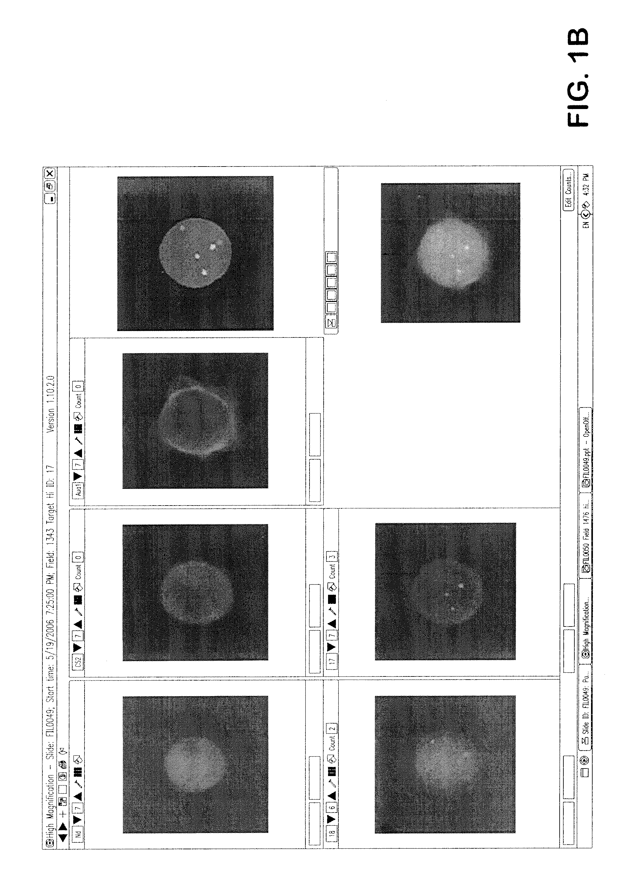
[0033]In one embodiment, an automated method of detecting circulating tumor cells in fluids obtained from patients, the tumor cells characterized by chemical and physical features of the cells, and generating information used to diagnose and / or monitor therapeutic progress comprising the steps of[0034]a) obtaining fluid from an individual;[0035]b) partially purifying tumor cells in fluid;[0036]c) contacting partially purified tumor cells with one or more distinguishable labeled probes directed to one or more characteristic features predetermined to identify tumor cells among normal cells feature under conditions to sufficiently affix labeled probe to withstand processing;[0037]d) analyzing said labeled cells with a high throughput robotic digital microscopy platform using[0038](i) a low magnification to locate said labeled cell followed by,[0039](ii) a high magnification for verification of cells bearing at least one said labeled probe; and from additional probes directed to charact...


PUM


Abstract

An automated, highly sensitive, specific and potentially quantitative detection method using an automated microscope for identifying and enumerating rare cancer cells in blood and other fluids.

Description

CROSS REFERENCE TO RELATED APPLICATIONS[0001]This application claims the benefit of priority of U.S. Provisional Application No. 61 / 002,836, filed Nov. 13, 2007. This reference and all additional references cited in this specification, and their references, are incorporated by reference herein in their entirety where appropriate for teachings of additional or alternative details, features, and / or technical background, and to the extent they do not contradict the specification below, in particular the definitions set forth below.BACKGROUND OF THE INVENTION[0002]1. Field of the Invention[0003]The present invention generally relates to an automated method for detecting circulating cancer cells in an individual. There is presented in one embodiment a method of assessing treatment protocols in the treatment of one or more cancers.[0004]2. Description of the Related Art[0005]Primary tumor cells presumably spread to other organs via blood and lymphatic circulation (1), and are probably she...

Claims


Application Information

Patent Timeline
IPC IPC(8): G06K9/00
CPCG06K9/00127G01N21/6458G06V20/69
Inventor BODMER, SIR WALTERTAFAS, TRIANTAFYLLOSKILPATRICK, MICHAELTSIPOURAS, PETROSWANG, XIUZHONGKIM, YOUNGMINTHOMAS, MICHAELSEPPO, ANTTINTOUROUPI, TRIANTAFYLLIAYU, CHANGHUACLIMER, CHARLES
Owner IKONISYS INC
Why Eureka
Try Eureka