Passive intraocular drug delivery devices and associated methods

Inactive Publication Date: 2009-06-04
ACIONT
View PDF5 Cites 23 Cited by
  • Summary
  • Abstract
  • Description
  • Claims
  • Application Information

AI Technical Summary

Benefits of technology

[0017]In another aspect of the present invention, a method for forming an active agent depot within the eye of a subject is provided. Such a method may include contacting a housing against an eye surface, the housing further including an active agent reservoir component located within the housing and configured to be fluidically coupled to a first ocular surface, and wherein the active agent reservoir contains an ionized active agent. The method may further include contacting a depot forming agent with the eye, the depot forming agent having a charge that is opposite in polarity as compared to the ionized active agent, and wherein the ionized active agent and the depot forming agent are configured to form a precipitate when contacted together at room to body temperature and at a pH of between about 4 to about 8 in an aqueous medium. Additionally, the method may include allowing sufficient time for the ionized active agent and the depot forming agent to passively diffuse into the eye and form an active agent depot. While contact of the active agent and the depot forming agent with the eye may be simultaneous, in one aspect the depot forming agent may be contacted with the eye prior to contacting the ocular lens-shaped housing with the eye. In another aspect, the depot forming agent may be contacted with the eye following contacting the ocular lens-shaped housing with the eye.

Problems solved by technology

Additionally, the device does not include an electrode.

Method used


Image

  • Passive intraocular drug delivery devices and associated methods
  • Passive intraocular drug delivery devices and associated methods
  • Passive intraocular drug delivery devices and associated methods

Examples


example 1

[0088]A 100 microliter / 30 G needle Hamilton syringe is used to administer a sub conjunctiva injection (n=2) of 20 microliters of 0.1M DSP at pH 7 into an eye of a New Zealand White Rabbit. The initial dose of DSP is 1.04 mg. After 4 hours from the time of the injection, the rabbit is euthanized and the eye is dissected and analyzed for the presence of DSP. The process is repeated for a total of 2 trials. The results are summarized in Table 1. About 23 micrograms of the approximate 1.04 mg subconjunctival injection of DSP are recovered from the whole eye after 4 hours. This illustrates the clearance effects of the transscleral pathway; in this case about 97.8 percent of the drug cleared the eye within four hours.

example 2

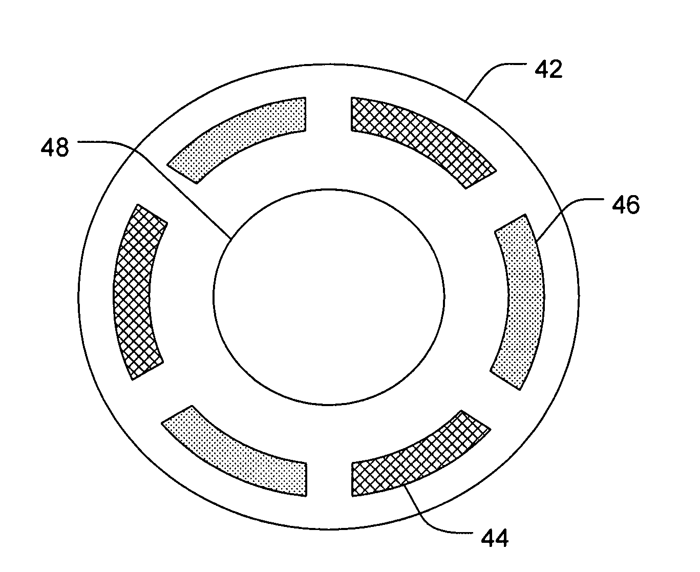
[0089]A six chambered annular lens-shaped housing is used to passively administer dexamethasone sodium phosphate (DSP) into the eye for immediate release. Chambers 1, 3, and 5 each contain a 2 mm Avalon Sponge hydrated with 25 μl of hydrogel and are further loaded with 6.47 mgs of 0.5 M DSP at pH 7 in each chamber. The housing is coupled to an eye of a New Zealand White Rabbit and depressurized with 0.2 cc of suction. The housing is maintained on the eye for 20 minutes. After 4 hours from the time the housing was applied to the eye, the rabbit is euthanized and the eye is dissected and analyzed for the presence of DSP. The process is repeated for a total of 4 trials. The results are summarized in Table 1.

example 3

[0090]A six chambered annular lens-shaped housing is used to passively administer DSP and a depot forming agent into the eye for sustained release. Each chamber of the housing contains a 2 mm Avalon Sponge hydrated with 25 μl of hydrogel. Chambers 1, 3, and 5 are further loaded with 6.47 mgs of 0.5 M dexamethasone sodium phosphate (DSP) at pH 7 in each chamber. Chambers 2, 4, and 6 are further loaded with 1.0 M CaCl2. The housing is coupled to an eye of a New Zealand White Rabbit and depressurized with 0.2 cc of suction. The housing is maintained on the eye for 20 minutes. After 4 hours from the time the housing was applied to the eye, the rabbit is euthanized and the eye is dissected and analyzed for the presence of DSP. The process is repeated for a total of 4 trials. The results are summarized in Table 1.

TABLE 1Example 3Example 1Example 2DSP-CaCl2DSP Total DrugDSP Total DrugTotal Drug(micrograms)(micrograms)(micrograms)Sclera10.8 ± 7.74.3 ± 1.046.7 ± 23.5Retina / Choroid 0.8 ± 0.21...


PUM


Abstract

The present invention provides methods and devices for the passive delivery of active agents into the eye of a subject. In one aspect, for example, a device for passively delivering an active agent into an eye of a subject may include a housing configured to conform to at least a portion of an eye surface, and a first reservoir component located within the housing and configured to be fluidically coupled to a first ocular surface, where the first reservoir contains an ionized active agent. The device may further include a second reservoir component located within the housing and configured to be fluidically coupled to a second ocular surface located adjacent to the first ocular surface, where the second reservoir component contains a depot forming agent having a charge that is opposite in polarity as compared to the ionized active agent. The ionized active agent and the depot forming agent are configured to form a precipitate when contacted together at room to body temperature and at a pH of between about 4 to about 8 in an aqueous medium. Additionally, the device does not include an electrode.

Description

FIELD OF THE INVENTION[0001]The present invention relates to systems, methods, and devices for the ocular delivery of an active agent into a subject's eye. Accordingly, the present invention involves the fields of chemistry, pharmaceutical sciences, and medicine, particularly ophthalmology.BACKGROUND OF THE INVENTION[0002]Posterior and intermediate eye diseases that require ocular drug delivery to prevent blindness include uveitis, bacterial and fungal endophthalmitis, age-related macular degeneration, viral retinitis, and diabetic retinopathy, among others. For example, the reported incidence of posterior uveitis is more than 100,000 people in the United States. If left untreated, uveitis leads to blindness. It is responsible for about 10 percent of all visual impairment in the U.S. and is the third leading cause of blindness worldwide.[0003]Treatments of intermediate and posterior uveitis are complicated by the inaccessibility of the posterior eye to topically applied medications....

Claims


Application Information

Patent Timeline
Patent Type & Authority Applications(United States)
IPC IPC(8): A61F9/00
CPCA61F9/0017
Inventor HIGUCHI, JOHN W.LI, S. KEVIN
Owner ACIONT
Why Eureka
Try Eureka