Medical Device Applications of Nanostructured Surfaces

a nanofiber and surface technology, applied in the field of nanofibers, can solve problems such as end users being susceptible to damage, and achieve the effects of enhancing surface properties, preventing/reducing bio-fouling, and increasing fluid flow

Inactive Publication Date: 2009-06-25
NANOSYS INC
View PDF60 Cites 41 Cited by
  • Summary
  • Abstract
  • Description
  • Claims
  • Application Information

AI Technical Summary

Benefits of technology

[0006]The embodiments of the current invention comprise various medical devices, such as clamps, valves, intracorporeal or extracorporeal devices (e.g., catheters), temporary or permanent implants, stents, vascular grafts, anastomotic devices, aneurysm repair devices, embolic devices, and implantable devices (e.g., orthopedic implants) and the like which comprise nanofiber enhanced surfaces. Such enhanced surfaces provide many enhanced attributes to the medical devices in, on, or within which they are used including, e.g., to prevent / reduce bio-fouling, increase fluid flow due to hydrophobicity, increase adhesion, biointegration, etc.
[0007]In a first aspect of the invention, a medical device is disclosed comprising a body structure having one or more surfaces having a plurality of nanostructured components associated therewith. The medical device may comprise an intracorporeal or extracorporeal device, a temporary or permanent implant, a stent, a vascular graft, an anastomotic device, an aneurysm repair device, an embolic device, an implantable device, a catheter, valve or other device which would benefit from a nanostructured surface according to the teachings of the present invention. The plurality of nanostructured components may comprise, for example, a plurality of nanofibers or nanowires. The plurality of nanostructured components enhance one or more of adhesion, non-adhesion, friction, patency or biointegration of the device with one or more tissue surfaces of a body of a patient depending on the particular application of the device. The nanofibers (or other nanostructured components) on the surfaces of the medical device can optionally be embedded in a slowly-soluble biocompatible polymer (or other) matrix to make the nanofiber surfaces more robust. The polymer matrix can protect most of the length of each nanofiber, leaving only the ends susceptible to damage. The generation of water soluble polymers can be accomplished in a number of different ways. For example, polymer chains can be formed in situ in a dilute aqueous solution primarily consisting of a monomer and an oxidizing agent. In this case, the polymer is actually created in the solution and subsequently spontaneously adsorbed onto the nanofiber surfaces as a uniform, ultra-thin film of between approximately 10 to greater than 250 angstroms in thickness, more preferably between 10 and 100 angstroms.
[0014]In another embodiment of the invention, a bioengineered scaffold device for providing a scaffold for nerve regeneration is disclosed which comprises a base membrane or matrix having a plurality of nanostructured components associated therewith. The membrane or matrix may be made from one or more of the following materials: natural or synthetic polymers, electrically conducting polymers, metals, alloys, ceramics, glass fabrics, or silicone. The plurality of nanostructured components may comprise, for example, a plurality of nanofibers or nanowires. The nanostructured surface of the membrane or matrix may be impregnated or bound with one or more drugs, cells, fibroblasts, nerve growth factors (NGF), cell seeding compounds, neurotrophic growth factors or genetically engineered cells producing such factors, VEGF, laminin or other drugs or substances to encourage axonal elongation and functional nerve performance.
[0019]Methods of use are also disclosed for treating patients with any one or more of the medical devices disclosed herein, which include, for example, a method of therapeutically treating a patient comprising contacting the patient with a medical device comprising a surface and plurality of nanofibers associated with the surface. Methods are disclosed for administering a drug compound to a body of a patient which comprises, for example, providing a drug-eluting device comprising at least one surface, a plurality of nanofibers associated with the surface, and a drug compound associated with the plurality of nanofibers; introducing the drug-eluting device into a body of a patient; and delivering the drug compound into the body of the patient. The drug-eluting device in one embodiment comprises a coronary stent, although any device which would benefit from local drug delivery at the site of disease (e.g., lesion) could be used in the methods of the invention. Where a coronary stent is used as the drug-eluting device, the drug compound may comprise paclitaxel or sirolimus, for example, or a variety of other medications including without limitation one or more of the following: anti-inflammatory immunomodulators such as Dexamethasone, M-prednisolone, Interferon, Leflunomide, Tacrolimus, Mizoribine, statins, Cyclosporine, Tranilast, and Biorest; antiproliferative compounds such as Taxol, Methotrexate, Actinomycin, Angiopeptin, Vincristine, Mitomycin, RestenASE, and PCNA ribozyme; migration inhibitors such as Batimastat, Prolyl hydroxylase inhibitors, Halofuginone, C-proteinase inhibitors, and Probucol; and compounds which promote healing and re-endothelialization such as VEGF, Estradiols, antibodies, NO donors, and BCP671. The drug compound may be adsorbed directly to the nanofiber surface of the drug-eluting device or otherwise associated with it via the use of one or more silane groups, linker molecules or other covalent, ionic, van der waals etc. attachment means. The nanofiber surface may be configured such that the drug compound elutes slowly over time. The plurality of nanofibers optionally are embedded in a biocompatible, non-thrombogenic polymer coating to provide enhanced durability to the nanofibers.

Problems solved by technology

The polymer matrix can protect most of the length of each nanofiber, leaving only the ends susceptible to damage.

Method used


Image

  • Medical Device Applications of Nanostructured Surfaces
  • Medical Device Applications of Nanostructured Surfaces
  • Medical Device Applications of Nanostructured Surfaces

Examples


Embodiment Construction

[0036]It should be appreciated that specific embodiments and illustrations herein of uses or devices, etc., which comprise nanofiber enhanced surface areas should not be construed as limiting. In other words, the current invention is illustrated by the descriptions herein, but is not constrained by individual specifics of the descriptions unless specifically stated. The embodiments are illustrative of various uses / applications of the enhanced surface area nanofiber surfaces and constructs thereof. Again, the enumeration of specific embodiments herein is not to be taken as limiting on other uses / applications which comprise the enhanced surface area nanofiber structures of the current invention.

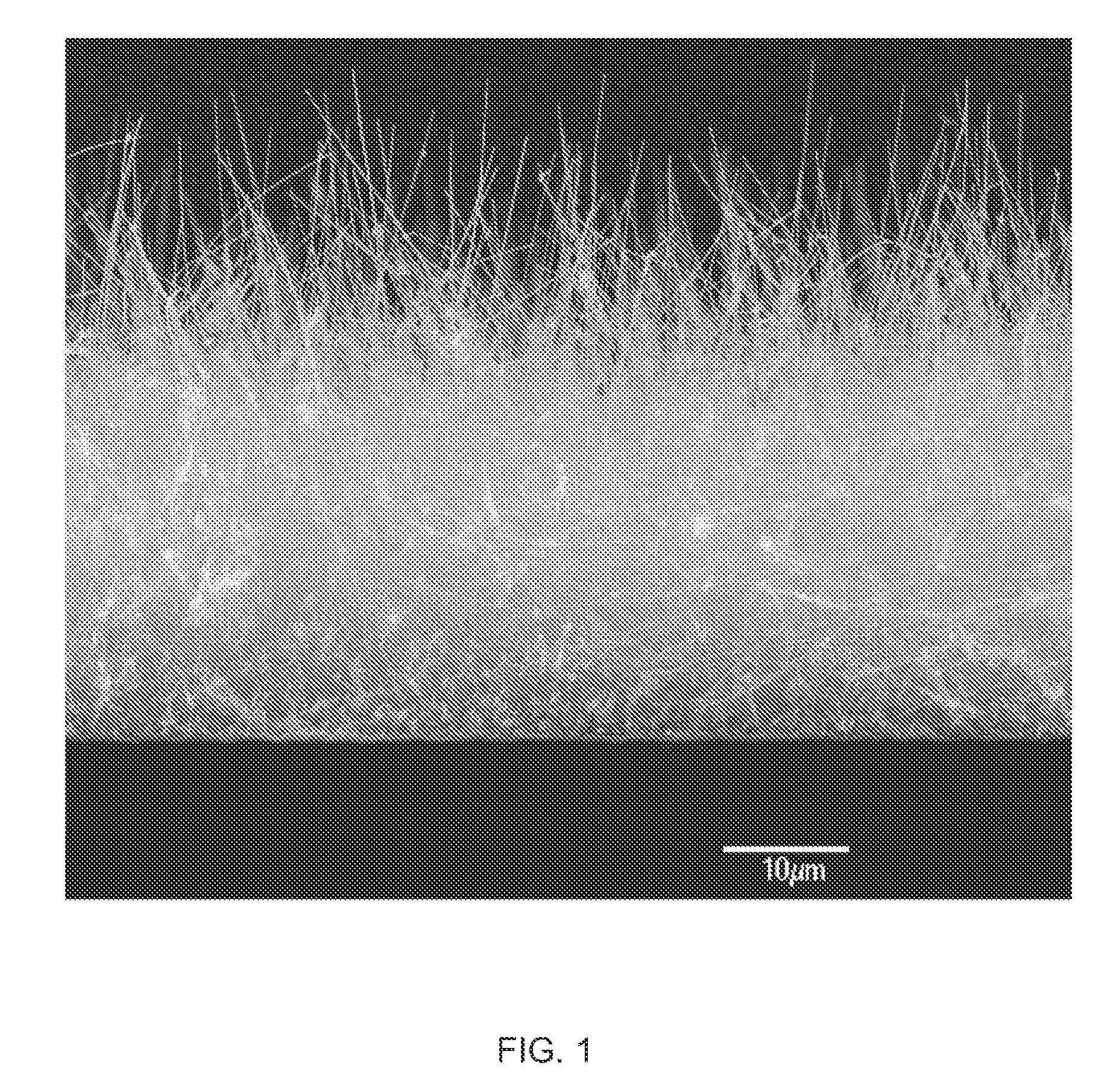
[0037]As seen in FIG. 1, the nanofibers optionally form a complex three-dimensional structure on the medical device surfaces to which they are applied. Again, it will be appreciated that in other embodiments of the invention, the nanofibers are more uniform in height, conformation, etc. The deg...


PUM

PropertyMeasurementUnit
thicknessaaaaaaaaaa
thicknessaaaaaaaaaa
thicknessaaaaaaaaaa

Abstract

This invention provides novel nanofiber enhanced surface area substrates and structures comprising such substrates for use in various medical devices, as well as methods and uses for such substrates and medical devices.

Description

CROSS-REFERENCE TO RELATED APPLICATIONS[0001]This application is a divisional of U.S. patent application Ser. No. 10 / 902,700, filed Jul. 29, 2004, which claims priority to U.S. Provisional Patent Application Ser. No. 60 / 549,711, filed Mar. 2, 2004. U.S. patent application Ser. No. 10 / 902,700, filed Jul. 29, 2004 also claims priority as a continuation-in-part application of U.S. patent application Ser. No. 10 / 828,100, filed Apr. 19, 2004, now U.S. Pat. No. 7,074,294, which is a continuation-in-part of U.S. patent application Ser. No. 10 / 661,381, filed Sep. 12, 2003, now U.S. Pat. No. 7,056,409, which claims priority to U.S. Provisional Patent Application No. 60 / 463,766, filed Apr. 17, 2003; and as a continuation-in-part of U.S. patent application Ser. No. 10 / 833,944, filed Apr. 27, 2004, which claims priority to U.S. Provisional Application Ser. No. 60 / 466,229, filed Apr. 28, 2003; and as a continuation-in-part application of U.S. patent application Ser. No. 10 / 840,794 filed May 5, 2...

Claims


Application Information

Patent Timeline
Patent Type & Authority Applications(United States)
IPC IPC(8): B32B5/16A61B17/08A61F2/06
CPCA61L31/14A61L2400/12Y10T428/259B82Y30/00A61L2400/18A61P31/04A61P35/00A61P41/00
Inventor DUBROW, ROBERT S.SLOAN, L. DOUGLAS
Owner NANOSYS INC
Why Eureka
Try Eureka