Proteolipid Membrane and Lipid Membrane Biosensor

a biosensor and lipid membrane technology, applied in the field of lipid membrane biosensors, can solve the problems of difficult assaying of lipids, affecting the structurally fragile environment, and often perturbing labels, and achieve the effect of increasing the number of samples/readings

Inactive Publication Date: 2009-07-30
X BODY
View PDF88 Cites 24 Cited by
  • Summary
  • Abstract
  • Description
  • Claims
  • Application Information

AI Technical Summary

Benefits of technology

[0005]The present invention provides biosensors in microtiter plate-based or microarray formats that allow for lipid and lipid-based assays with much higher sample number / readings per unit time. Currently, a typical assay for a single binding interaction event with a liposome SPR sensor on a single instrument takes 20-30 minutes. See, e.g., Baird et al., Analyt. Biochem. 2002 310:93-99. In this amount of time, the present invention could make readings for 4 to 6×384-well sample biosensor plates on a single instrument. Advantageously, labels are not required for detection in the instant invention.

Problems solved by technology

It can be difficult to assay lipids with conventional biosensors such as Biacore's X, 2000, 3000, T100, S51 and C systems or label-requiring techniques such as fluorescence-based approaches because labels do not work well in non-polar environments having various issues with quenching or excitation as known to those practiced in the art.
In addition, labels often perturb systems they are used to study lipids in unpredictable and more importantly in unwanted ways.
Flow based systems such as Biacore perturb the structurally fragile environment required to maintain and make measurements in non-polar environments.
In addition, SPR flow formats provide only very low sample throughput which can add considerably to the time to develop proper environments, allow proper protein attachment, assembly & folding, and increase the amount of time to finally test for any interactions with a protein should it become properly attached and folded in the flow environment.

Method used


Image

  • Proteolipid Membrane and Lipid Membrane Biosensor
  • Proteolipid Membrane and Lipid Membrane Biosensor
  • Proteolipid Membrane and Lipid Membrane Biosensor

Examples


example 1

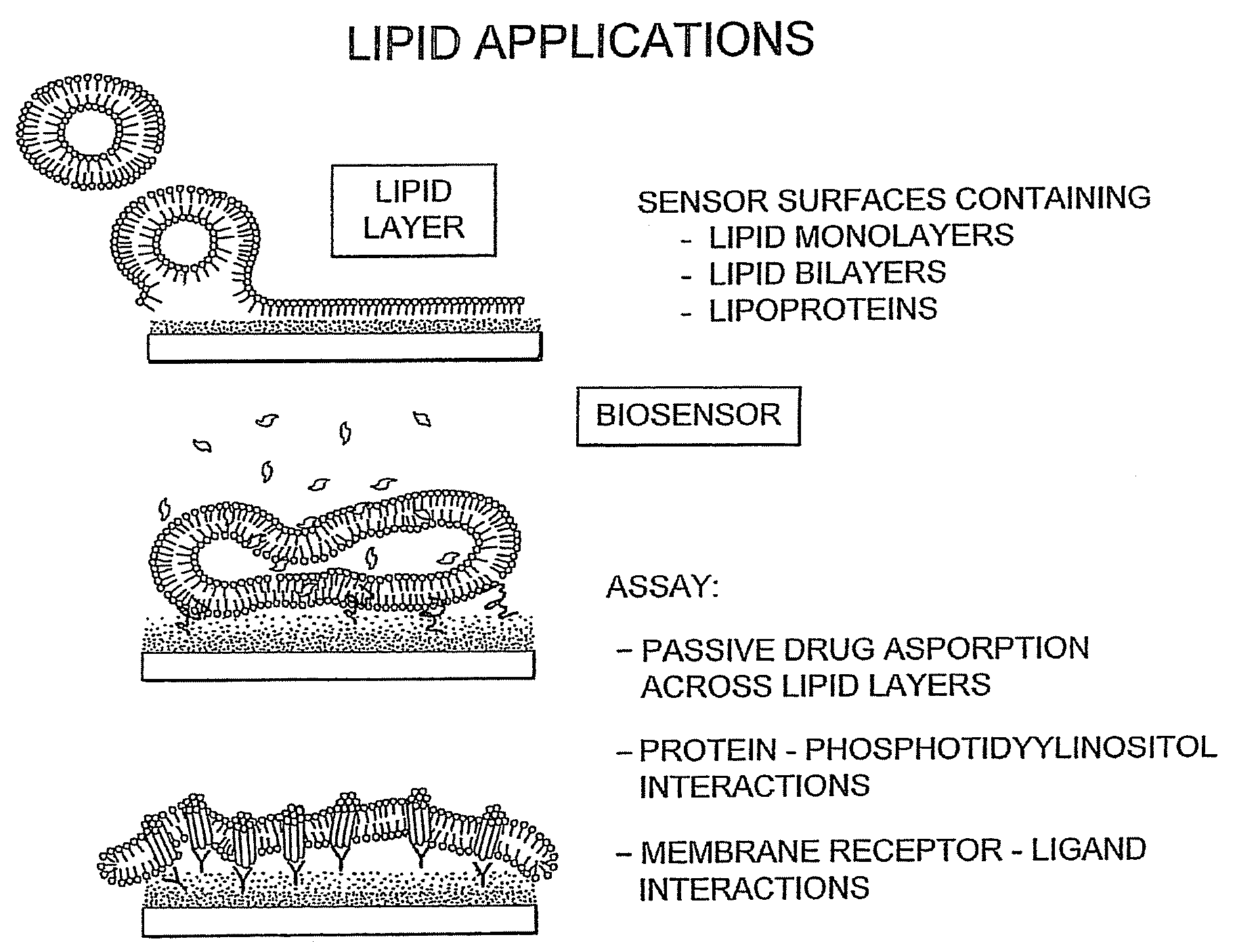
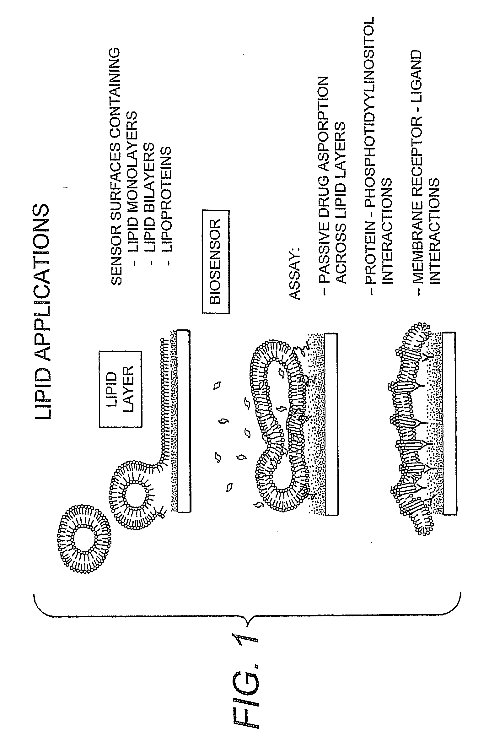
Stable Hydrophobic Biosensor Surfaces

[0042]A stable hydrophobic calorimetric resonant biosensor surface can be prepared by injecting repel silane (Amersham Biosciences 17-1332-01) into wells comprising a calorimetric resonant biosensor TiO surface, for example a 96-well plate. Incubate for 7 minutes. Inject 90 ul hexane (anhydrous) (Sigma 227064) into each well. Aspirate 90 ul of silane mixture with a multi-channel pipette. Inject 90 ul hexane (2nd injection) into wells. Aspirate 90 ul of silane mixture (2nd aspiration) with a multi-channel pipette. Inject 90 ul hexane (3rd injection) into wells. Aspirate all remaining solution in wells using an 8-channel stainless steel manifold connected to a vacuum pump. Allow the biosensor to cure in air after final aspiration. Wash the plate 2× with 100 μL PBS, then fill plate with 100 μL PBS. Sonicate the plate for 5-10 sec moving the plate back and forth in the sonication bath to disperse energy evenly across plate. Dry the backside of plate ...

example 2

Hydrophobic Silane Sensor Treatment Poly-Phe-Lys Deposition Test for Hydrophobicity

[0043]

TABLE 1Plate 1Plate 2Plate 3Plate 4Plate 5PPLShiftSD% CVShiftSD% CVShiftSD% CVShiftSD% CVShiftSD% CVNO4.00.13.54.10.13.44.00.13.34.10.13.34.10.12.9HEXANE4.20.12.54.20.12.94.00.12.84.10.12.54.10.12.8REPEL4.80.612.64.80.612.14.60.25.06.01.626.46.81.623.1N for each reported number is 32 wells

[0044]A colorimetric resonant biosensor surface was treated with repel silane (2% w / v in octamethylcyclotetrasiloxane) by incubating 50 μl of repel silane for a 7 minute treatment in the wells of a microtiter biosensor plate. The silane solution was aspirated and the surface washed 3× with 90 μl of hexanes. The microtiter biosensor plate was cured for 5 minutes after final hexanes wash by allowing the plate to dry. The plate was washed 3× with PBS pH 7.4. PPL was deposited onto the silane treated biosensor surface. 40 μl of 0.1 mg / ml PPL in 10 mM sodium phosphate pH 9.05 was added to each 6 mm diameter well of ...

example 3

Repel Silane Treatment to Improve PPL Binding

[0051]A biosensor plate was O2 plasma treated prior to repel treatment. A 7 minute incubation of 50 μl repel silane treatment was performed in the 6 mm diameter wells of biosensor plates. The remaining silane was aspirated and the biosensor was washed 3× with 90 μl of hexane. The biosensor plate was cured for 5 minutes after the final hexane wash. The biosensor plate was washed 3× with PBS pH 7.4. 0.1 mg / ml PPL solutions were prepared in 10 mM NaH2PO3 buffers at pHs of 6.0, 7.4, 9.0, and 10.1, these buffers were prepared by adjusting an 11 mM NaH2PO3 pH 4.0 buffer with 1 M NaOH. 40 ml of the PPL solutions were added to the wells of the biosensor plate and dried overnight at RT. The biosensor plate was washed 3× with PBS. FIG. 6 shows the results of optimizing the hydrophobic coating to improve the capture of the model protein PPL.


PUM

PropertyMeasurementUnit
thicknessaaaaaaaaaa
diameteraaaaaaaaaa
pHaaaaaaaaaa

Abstract

The invention provides compositions and methods for detection of interaction of molecules.

Description

PRIORITY INFORMATION[0001]This application is a divisional of U.S. Ser. No. 11 / 403,128, which was filed on Apr. 12, 2006, which claims the benefit of U.S. Ser. No. 60 / 670,524, filed on Apr. 12, 2005, which are incorporated herein by reference in their entirety.BACKGROUND OF THE INVENTION[0002]It can be difficult to assay lipids with conventional biosensors such as Biacore's X, 2000, 3000, T100, S51 and C systems or label-requiring techniques such as fluorescence-based approaches because labels do not work well in non-polar environments having various issues with quenching or excitation as known to those practiced in the art. In addition, labels often perturb systems they are used to study lipids in unpredictable and more importantly in unwanted ways. Flow based systems such as Biacore perturb the structurally fragile environment required to maintain and make measurements in non-polar environments. In addition, SPR flow formats provide only very low sample throughput which can add co...

Claims


Application Information

Patent Timeline
Patent Type & Authority Applications(United States)
IPC IPC(8): C40B30/04G01N21/55G01N21/59G01N21/41
CPCG01N33/5432G01N33/92G01N33/551G01N33/54373
Inventor BAIRD, CHERYL L.WANG, GUO BINLAING, LANCEJOGIKALMATH, GANGADHARGERSTENMAIER, JOHN
Owner X BODY
Why Eureka
Try Eureka