Radiation image radiographing system

a radiographing system and radiographing technology, applied in medical science, diagnostics, electrical equipment, etc., to achieve the effects of reducing the sending time of radiograph image information for display, and enhancing image verification efficiency

Inactive Publication Date: 2009-08-06
KONICA MINOLTA MEDICAL & GRAPHICS INC
View PDF1 Cites 10 Cited by
  • Summary
  • Abstract
  • Description
  • Claims
  • Application Information

AI Technical Summary

Benefits of technology

[0038]According to the invention described in Claim 1, the console for operating the radiographic image detecting apparatus at the time of radiographing operation is normally positioned close to the X-ray room for this radiographing operation to ensure operation convenience, whereby this X-ray room is managed. To be more specific, association is established between the radiographic image detecting apparatus and patient by in the console positioned close to the X-ray room for this radiographing operation to ensure the easiest operation by the radiology technician in charge of radiographing operation, and radiographic image information is provided by the console having established this association. Further, the radiographic image information sent to the console is displayed on the display section of the console. This allows the radiology technician to determine suitability of radiographing operation and need of repeating the radiographing operation immediately after the first radiographing operation, with the result that image verification efficiency is enhanced. This has the effect of sufficiently demonstrating the advantages of the radiographic image detecting apparatus that does not require time from radiographing to reading.
[0039]According to the invention described in Claim 2, the radiographic image information for display can be sent to the console. By checking the radiographic image information for display appearing on the display section of the console, the radiology technician can determine suitability of radiographing operation, and the need of repeating the radiographing operation immediately after the first radiographing operation. Thus, image verification efficiency is enhanced.
[0040]According to the invention described in Claim 3, the radiographic image information for display is the reduced size image information whose information amount is smaller than that of the original image information. The sending time of the radiographic image information for display is reduced by sending only such reduced-size image information to the console, and quick display of the radiographic image information on the console can be achieved after radiographic imaging. This allows the radiology technician to determine suitability of radiographing operation and the need of repeating the radiographing operation immediately after the first radiographing operation. Thus, image verification efficiency is enhanced.
[0041]According to the invention described in Claim 4, a cassette type FPD is used as the radiographic image detecting apparatus. This arrangement provides the advantage of excellent transportability independently of the place where radiographing operation is performed.
[0042]According to the invention described in Claim 5, the radiographic image information obtained from the result of detection associated with the patient information is sent to the console associated with the aforementioned radiographic image detecting apparatus that has captured the image of the aforementioned patient last. For example, when radiographing operation has been repeated several times for one patient by moving between X-ray rooms, the radiographic image information for all the radiographing operations can be obtained at one console which is located closest to the X-ray room wherein the last radiographing operation has been performed. The radiographic image information sent to the console is displayed on the display section of the console. Accordingly, even when a plurality of consoles are arranged through one network and radiographing operation has been repeated several times for one patient by moving between X-ray rooms. This allows the radiology technician to determine suitability of radiographing operation and need of repeating the radiographing operation, without moving away from the console wherein the aforementioned last radiographing operation has been performed, with the result that image verification efficiency is enhanced.
[0043]According to the invention described in Claim 6, selecting a patent from the patient list makes it possible to get the radiographic image information based on the result of detection associated with the patient information, using the console wherein the patient has been selected. The radiographic image information sent to the console is displayed on the display section of the console. Accordingly, even when a plurality of consoles are arranged through one network and radiographing operation has been repeated several times for one patient by moving between X-ray rooms, the radiology technician can determine suitability of all radiographing operations for this particular patient and the need of repeating the radiographing operations, using a simple means. This arrangement leads to enhanced image verification efficiency.

Problems solved by technology

However, for recent development of the communication network, limited space and other various reasons, one console is often connected with a plurality of FPDs.

Method used


Image

  • Radiation image radiographing system
  • Radiation image radiographing system
  • Radiation image radiographing system

Examples


embodiment 1

[0069]The following describes the first embodiment of the present invention with reference to FIG. 1 through FIG. 4:

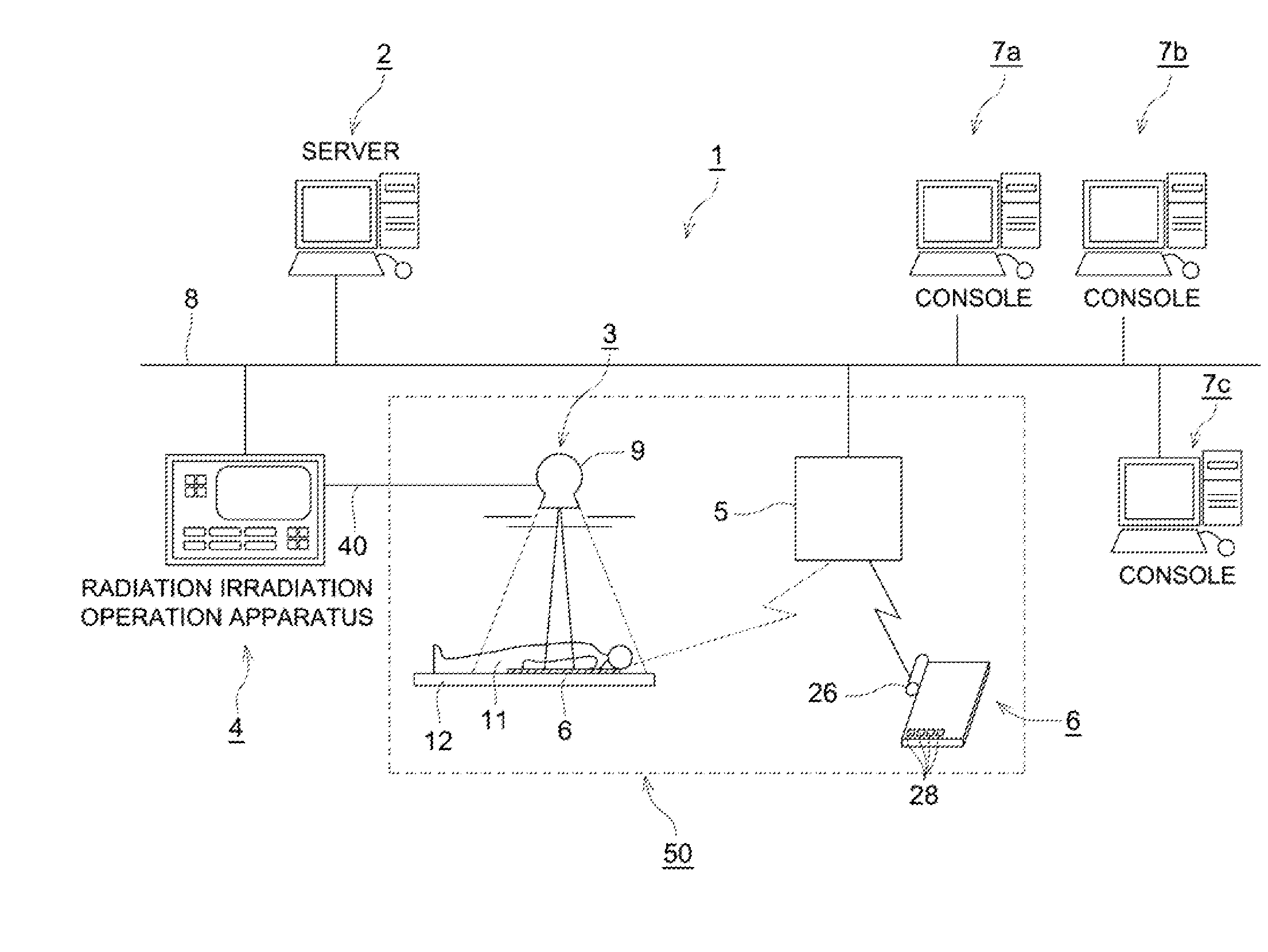
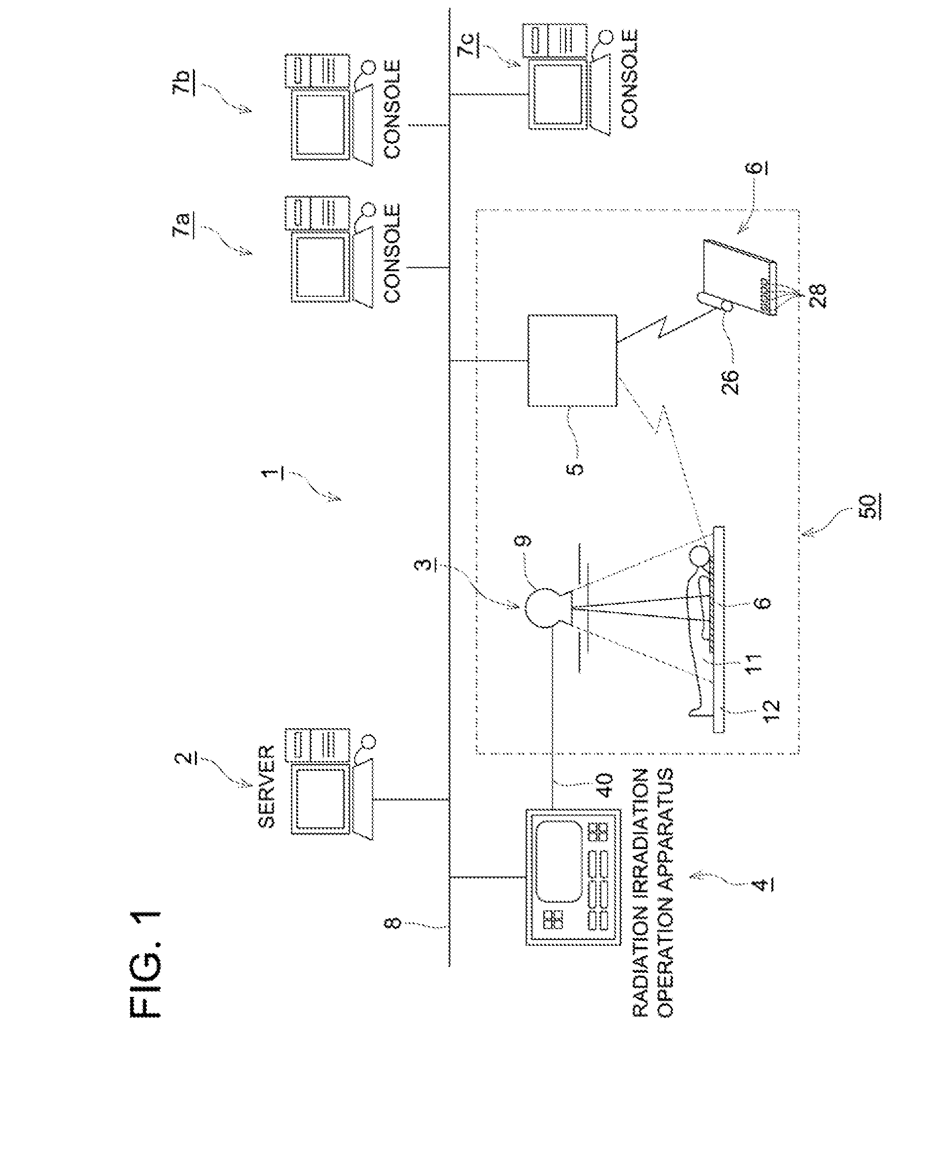
[0070]FIG. 1 is a diagram representing the schematic structure illustrating an embodiment of the radiation image radiographing system 1 applied with the radiographic image detecting apparatus of the present invention.

[0071]The radiation image radiographing system 1 of the present embodiment uses radiation such as X-rays to perform an radiographing operation. As shown in FIG. 1, a server 2 for managing the information on radiographic imaging operation, a radiation irradiation operation apparatus 4 for performing operation relating to irradiation with radiation, a base station 5 for performing communication by a radio communication method such as radio LAN (Local Area Network), and consoles 7a, 7b and 7c for managing the X-ray room 50 and operating the radiographic image detecting apparatus 6 installed in the X-ray room 50 are connected with one another via the network 8...


PUM


Abstract

A radiation image radiographing system using a combination of a plurality of consoles and a radiographic image detecting apparatus characterized by enhanced image verification efficiency. This radiation image radiographing system includes a radiographic image detecting apparatus for detecting an applied radiation, a plurality of consoles provided with a communication section for communicating with an external apparatus, the consoles being used to operate the radiographic image detecting apparatus, a control section as an association device for establishing associations between the radiographic image detecting apparatus and the console for operating the radiographic image detecting apparatus during radiographing operation, and a control section as an image information transmission device for sending the radiographic image information obtained based on the detection result of the radiographic image detecting apparatus, to at least the console associated by the control section, wherein the console has a display section for displaying the radiographic image information.

Description

FIELD OF THE INVENTION[0001]The present invention relates to a radiation image radiographing system, particularly a radiation image radiographing system for capturing a radiographic image using a plurality of consoles and radiographic image detecting apparatus.BACKGROUND OF THE INVENTION[0002]In the conventional art of medical diagnosis, a subject is irradiated with radiation such as X rays, and a radiographic image is obtained by detection of the distribution of the intensity of radiation having passed through the subject. This radiographic image has been employed widely in medical diagnosis. In recent years, a radiation image radiographing system has been developed, which uses an FPD (Flat Panel Detector) as a radiographic image detecting apparatus wherein a radiation is detected as radiographic image information at the time of photographing and is converted into an electric signal.[0003]One of these radiation image radiographing systems is linked with a predetermined console such...

Claims


Application Information

Patent Timeline
Patent Type & Authority Applications(United States)
IPC IPC(8): H05G1/64
CPCA61B6/00A61B6/4494A61B6/563
Inventor OHARA, HIROMU
Owner KONICA MINOLTA MEDICAL & GRAPHICS INC
Why Eureka
Try Eureka