Bioabsorbable Stent Having a Radiopaque Marker

a bioabsorbable, radiopaque technology, applied in the field of stents, can solve the problems of irritating the surrounding tissue, affecting the effect of the stent, and the difficulty of subsequent procedures, so as to avoid potential complications, avoid dislocation and embolism, and avoid the effect of increasing the risk of embolism

Inactive Publication Date: 2009-08-13
MEDTRONIC VASCULAR INC
View PDF16 Cites 105 Cited by
  • Summary
  • Abstract
  • Description
  • Claims
  • Application Information

AI Technical Summary

Benefits of technology

[0038]Embodiments of the present invention relate to a bioabsorbable stent having one or more radiopaque markers that are visible to a physician viewing, for example, an X-ray fluoroscopy device while deploying and / or positioning the stent into the body vessel. Radiopaque markers are generally secured to the proximal and / or distal ends of the stent extending outwardly from one or more peaks or troughs of undulating bands of the stent body. Embodiments of the present invention are directed to underlying stent structures that allow the one or more markers to endothelialize. Because the stent bioresorbs or breaks down and the marker does not, it is important that the marker remains fixed and stable during bioresorption of the stent body. By controlling dissolution of an area of the stent near the marker, the marker may endothelialize and is therefore prevented from dislodging and embolizing. Thus, the bioabsorbable stent may be made more radiopaque without increasing the risk of embolism caused by the dislodgement of the marker.
[0039]Dissolution of the biodegradable stent material or portion holding the marker in place (hereinafter referred to as marker support) is controlled or slowed so that it will remain intact for a sufficient time to allow for marker endothelialization. The term “endothelialization” is meant to describe the process in which a foreign object, such as the marker in embodiments of the present invention, becomes incorporated into the walls of the lumen by tissue ingrowth or encapsulation. Thus, in other words, dissolution of the marker support is controlled so that the marker is held against the vessel wall long enough to endothelialize. As part of the vessel wall, the marker is stable and will not migrate downstream and thus avoids causing potential complications. Dissolution of the marker support must be controlled or slowed for a sufficient time to allow for endothelialization to occur, approximately three to six weeks. The biodegradable body portion of the stent has a first dissolution rate and the marker support has a second dissolution rate. The second dissolution rate is slower than the first dissolution rate. In particular, the second dissolution rate is approximately 30-100% slower than the first dissolution rate in order to allow the radiopaque marker to endothelialize. The controlled dissolution of the marker support may be accomplished via one or more mechanisms that include the following: increasing the cross-sectional thickness of the marker support, passivating or oxidizing the marker support, utilizing a different, slower absorbing material for marker support, utilizing a bioabsorbable polymeric coating on the marker support, anodically protecting the marker support with a sacrificial anode, and any other suitable means of slowing absorption or corrosion in the region that secures the marker.
[0040]In an embodiment of the present invention, rather than delay dissolution of the entire stent in order to allow the radiopaque marker to endothelialize, it is desirable to selectively control or delay dissolution of only the stent material securing the marker. Selectively controlling dissolution of only the marker support material allows the remainder of the stent body to be absorbed in a desired amount of time and avoids the risk of the stent body becoming encapsulated and covered with endothelium tissues. In other words, if dissolution of the entire stent was controlled or delayed in order to allow the radiopaque marker to endothelialize, the stent body may also endothelialize and thus may not break down as desired. The biodegradable stent body must be in contact with a body fluid such as blood in order for the stent to corrode or be absorbed into the body as desired. Thus, selectively controlling dissolution of only the marker support avoids the undesirable endothelialization of the stent body.

Problems solved by technology

Permanent stents, over time, may become encapsulated and covered with endothelium tissues, for example, in cardiovascular applications, causing irritation to the surrounding tissue.
Further, if an additional interventional procedure is ever warranted, a previously permanently implanted stent may make it more difficult to perform the subsequent procedure.
However, there are disadvantages and limitations associated with the use of bioabsorbable or biodegradable stents.
Limitations arise in controlling the breakdown of the bioabsorbable materials from which such stents are made, as in, preventing the material from breaking down too quickly or too slowly.
If the material is absorbed too quickly, the stent will not provide sufficient time for the vessel to heal, or if absorbed too slowly, the attendant disadvantages of permanently implanted stents may arise.
Such magnesium stents, however, are not very radiopaque because magnesium does not show up well under fluoroscopy.
However, a problem that arises using a radiopaque marker with a biodegradable stent is a risk of embolism caused by the dislodgement of the marker that can then move downstream, which may occur when the biodegradable stent is absorbed by the body, but the marker is not.
Once the stent biodegrades, the marker may embolize and block the coronary arteries, or migrate further downstream, causing additional complications.

Method used


Image

  • Bioabsorbable Stent Having a Radiopaque Marker
  • Bioabsorbable Stent Having a Radiopaque Marker
  • Bioabsorbable Stent Having a Radiopaque Marker

Examples


Embodiment Construction

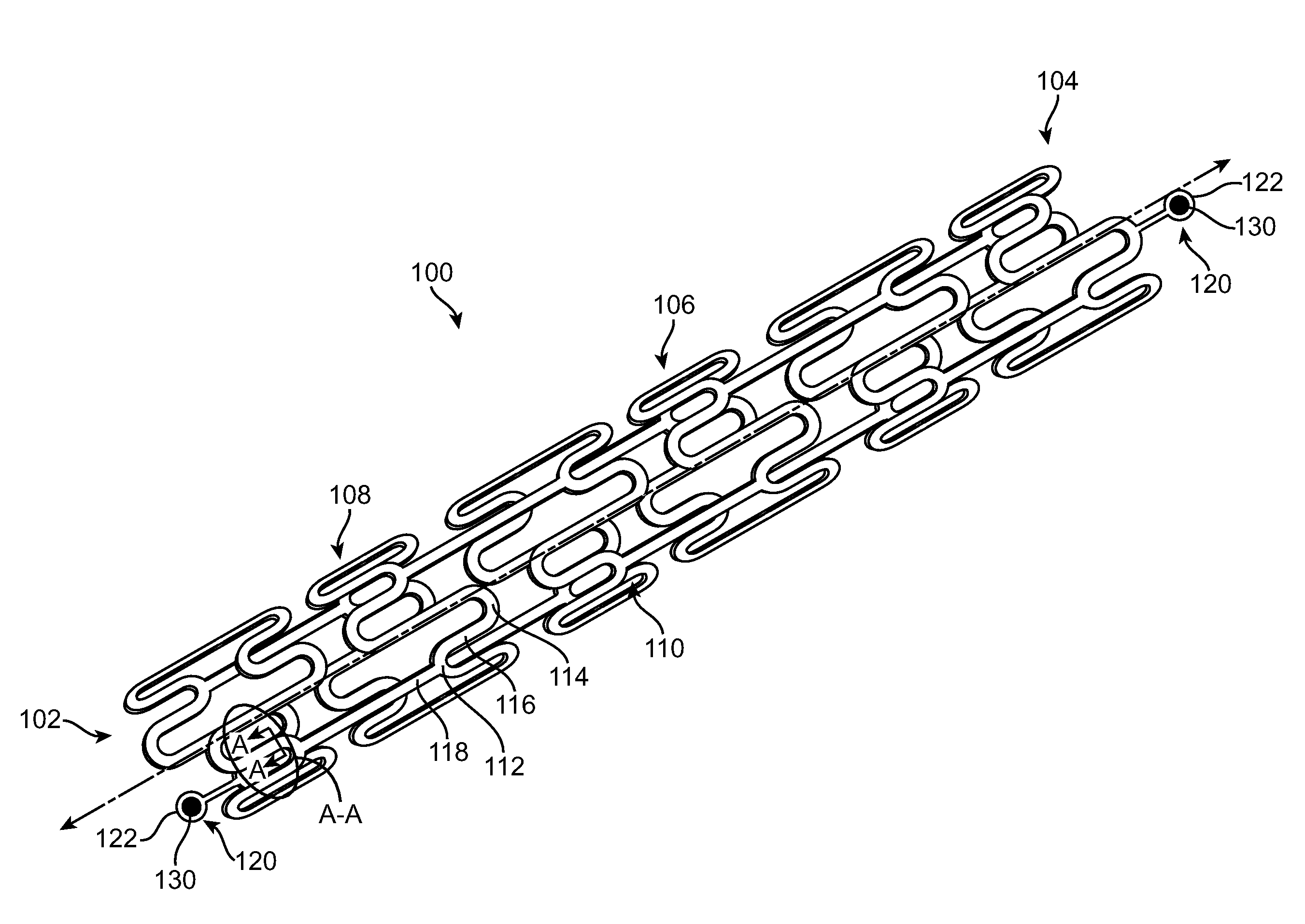
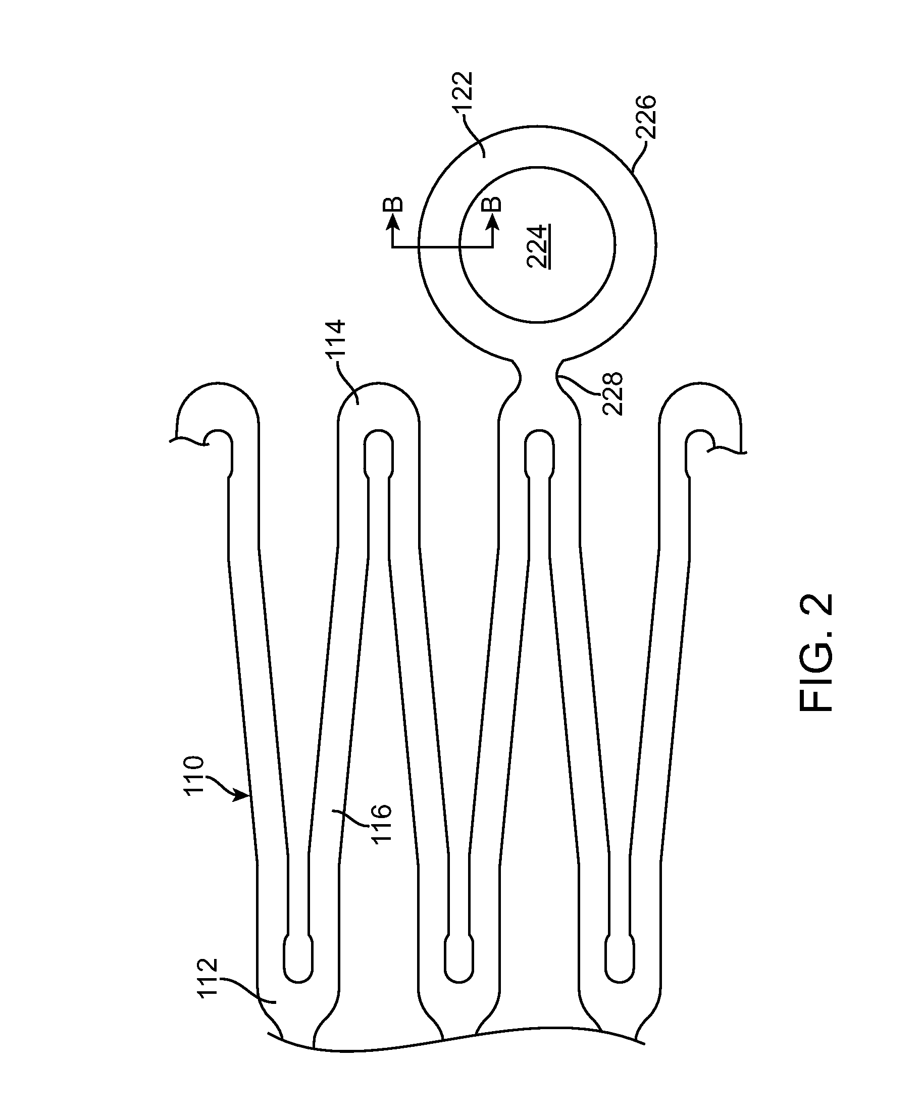
[0036]Specific embodiments of the present invention are now described with reference to the figures, wherein like reference numbers indicate identical or functionally similar elements. The terms “distal” and “proximal” are used in the following description with respect to a position or direction relative to the treating clinician. “Distal” or “distally” are a position distant from or in a direction away from the clinician. “Proximal” and “proximally” are a position near or in a direction toward the clinician.

[0037]The following detailed description is merely exemplary in nature and is not intended to limit the invention or the application and uses of the invention. Although the description of the invention is in the context of treatment of blood vessels such as the coronary, carotid and renal arteries, the invention may also be used in any other body passageways where it is deemed useful. Furthermore, there is no intention to be bound by any expressed or implied theory presented in ...


PUM

PropertyMeasurementUnit
timeaaaaaaaaaa
thicknessaaaaaaaaaa
radiopaqueaaaaaaaaaa

Abstract

A bioabsorbable stent includes one or more radiopaque markers. The stent body may include a generally cylindrical body portion and a marker support for receiving the one or more marker(s). The marker support may be connected to an end of the body portion, or may be an integral portion of the body portion. By selectively controlling dissolution of the biodegradable material of the marker support, the marker support will remain intact for a sufficient time to allow for the marker to endothelialize and therefore prevent the marker from dislodging and embolizing. The controlled dissolution may be accomplished via one or more of the following mechanisms, including increasing the cross-sectional thickness of the marker support, passivating or oxidizing the marker support, utilizing a different, slower absorbing material for the marker support, utilizing a bioabsorbable polymeric coating on the marker support, or protecting the marker support with a sacrificial anode.

Description

FIELD OF THE INVENTION[0001]The invention relates generally to temporary endoluminal prostheses for placement in a body lumen, and more particularly to stents that are bioabsorbable.BACKGROUND OF THE INVENTION[0002]A wide range of medical treatments exist that utilize “endoluminal prostheses.” As used herein, endoluminal prostheses is intended to cover medical devices that are adapted for temporary or permanent implantation within a body lumen, including both naturally occurring and artificially made lumens, such as without limitation: arteries, whether located within the coronary, mesentery, peripheral, or cerebral vasculature; veins; gastrointestinal tract; biliary tract; urethra; trachea; hepatic shunts; and fallopian tubes.[0003]Accordingly, a wide assortment of endoluminal prostheses have been developed, each providing a uniquely beneficial structure to modify the mechanics of the targeted lumen wall. For example, stent prostheses are known for implantation within body lumens t...

Claims


Application Information

Patent Timeline
Patent Type & Authority Applications(United States)
IPC IPC(8): A61F2/82
CPCA61F2/915A61F2210/0004A61F2230/0013A61F2250/0036A61F2250/0098A61F2250/003
Inventor ALLEN, JEFFREYBIRDSALL, MATTHEW J.
Owner MEDTRONIC VASCULAR INC
Why Eureka
Try Eureka