
Aspirator and method for pneumostoma management

a pneumoma and aspirator technology, applied in the direction of respirators, catheters, wound drains, etc., can solve the problems of increased exhalation work, hyperinflation of the lung, and significant underdiagnosis of psoriasis, and achieve the effect of stabilizing the artificial apertur

Inactive Publication Date: 2009-08-20
PORTAERO
View PDF101 Cites 24 Cited by
  • Summary
  • Abstract
  • Description
  • Claims
  • Application Information

AI Technical Summary

Benefits of technology

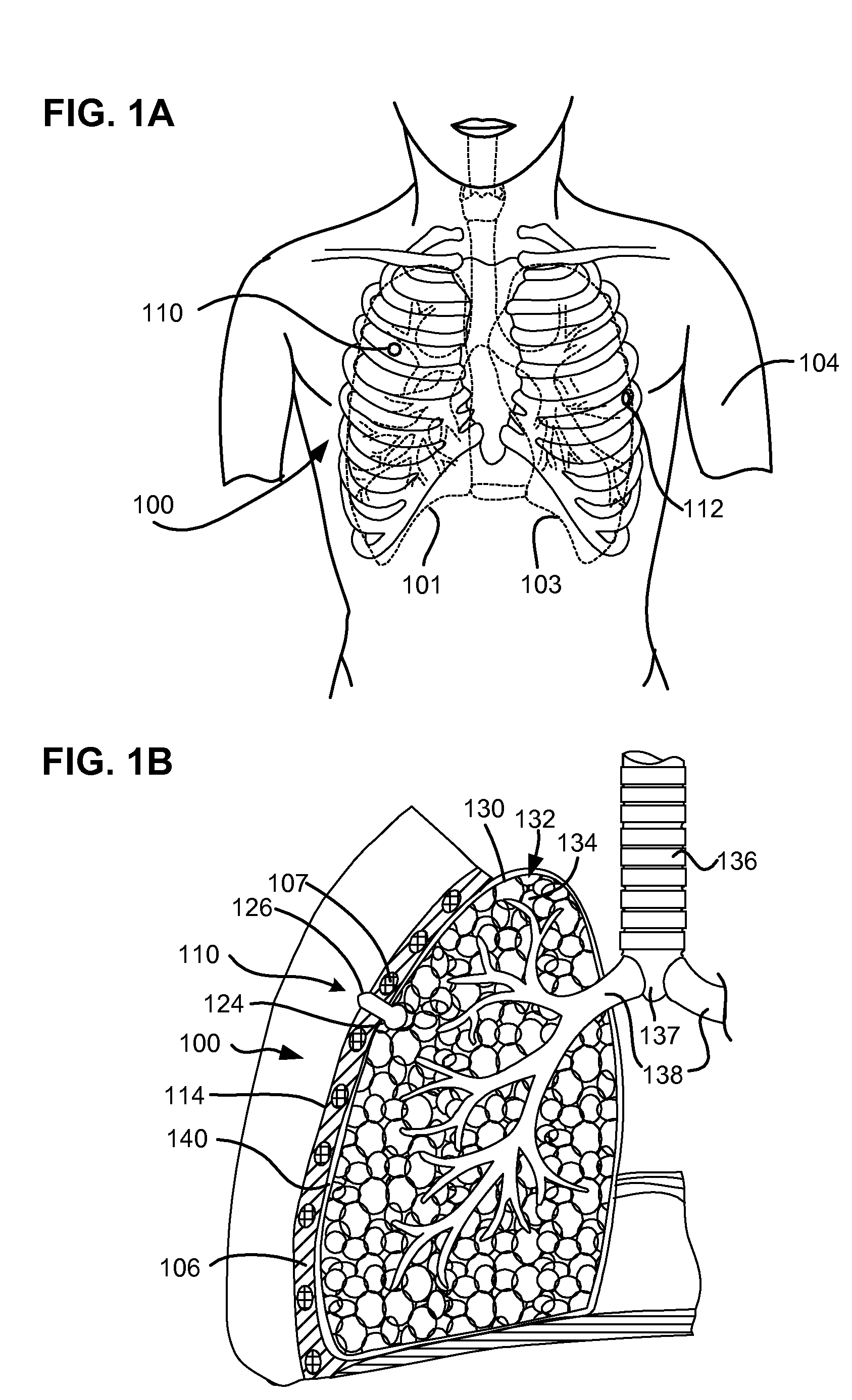
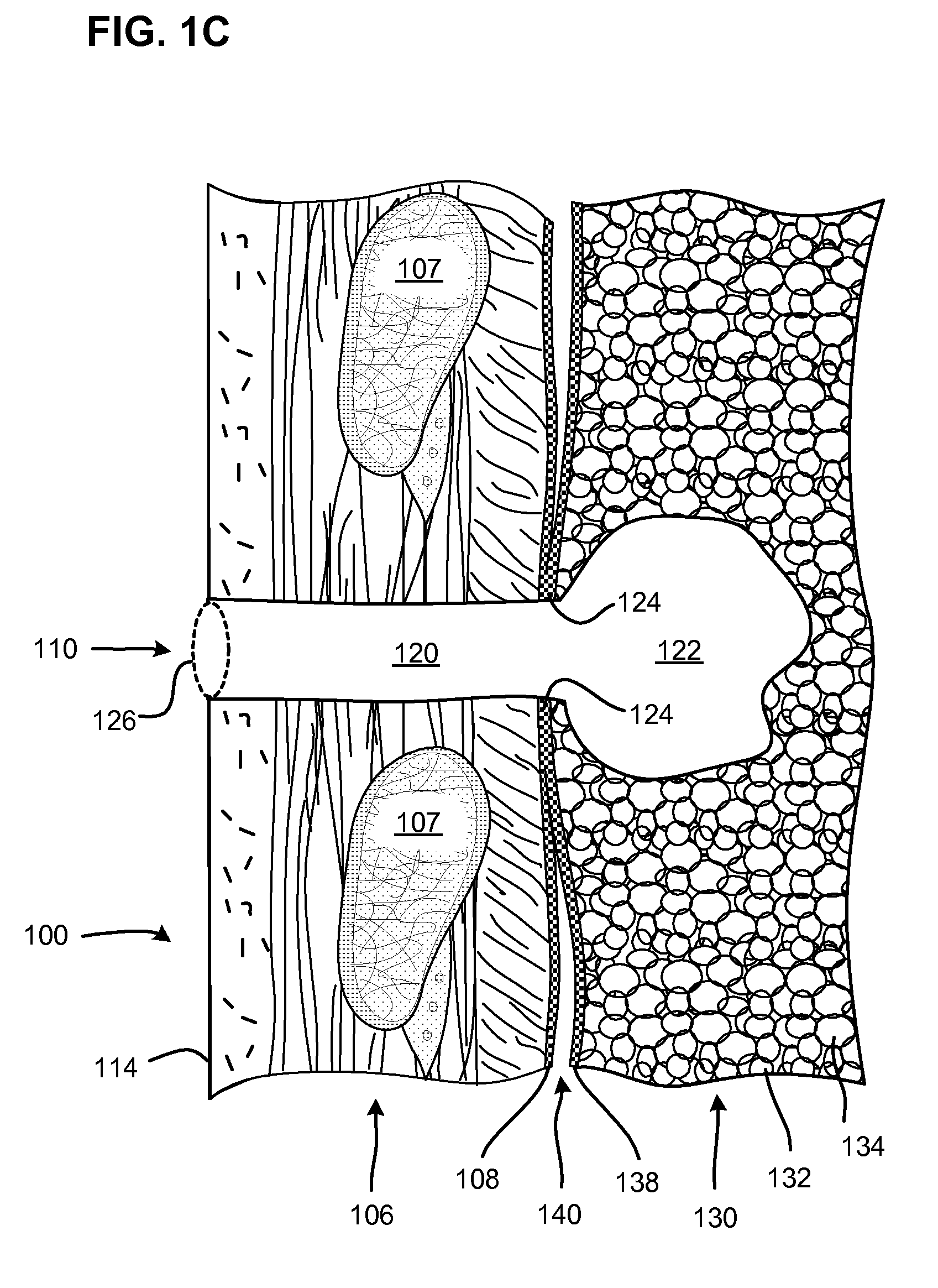
[0036]In view of the disadvantages of the state of the art, Applicants have developed a method for treating COPD in which an artificial passageway is made through the chest wall into the lung. An anastomosis is formed between the artificial passageway and the lung by creating a pleurodesis between the visceral and parietal membranes surrounding the passageway as it enters the lung. The pleurodesis prevents air from entering the pleural cavity and causing a pneumothorax (deflation of the lung due to air pressure in the pleural cavity). The pleurodesis is stabilized by a fibrotic healing response between the membranes. The artificial passageway through the chest wall also becomes epithelialized. The result is a stable artificial aperture through the chest wall which communicates with the parenchymal tissue of the lung.
[0037]The stable artificial aperture into the lung through the chest is referred to herein as a pneumostoma. A pneumostoma provides an extra pathway that allows air to exit the lung while bypassing the natural airways which have been impaired by COPD and emphysema. By providing this ventilation bypass, the pneumostoma allows the stale air trapped in the lung to escape from the lung thereby shrinking the lung (reducing hyperinflation). By shrinking the lung, the ventilation bypass reduces breathing effort (reducing dyspnea), allows more fresh air to be drawn in through the natural airways and increases the effectiveness of all of the tissues of the lung for gas exchange. Increasing the effectiveness of gas exchange allows for increased absorption of oxygen into the bloodstream and also increased removal of carbon dioxide. Reducing the amount of carbon dioxide retained in the lung reduces hypercapnia which also reduces dyspnea. The pneumostoma thereby achieves the advantages of lung volume reduction surgery without surgically removing or sealing off a portion of the lung.
[0038]In some situations, mucus / discharge and / or foreign matter may accumulate in the pneumostoma or a medical device implanted in the pneumostoma. An aspirator and methods of use in accordance with embodiments of the present invention are desirable and useful to remove the mucus / discharge and / or foreign matter in order to maintain the patency of the pneumostoma and prevent infection.
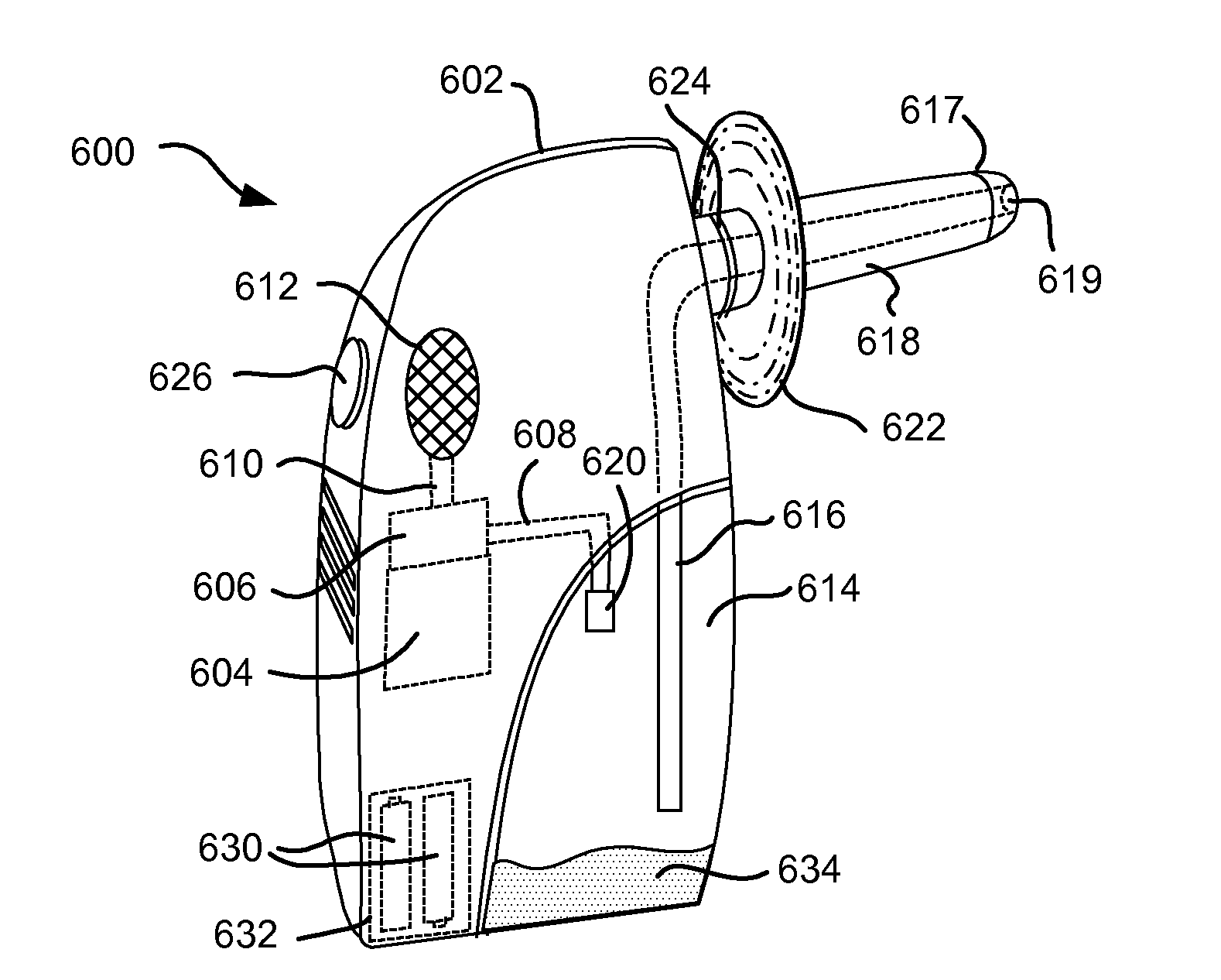
[0040]In accordance with one embodiment, the present invention provides a pneumostoma management system which includes a partially-implantable pneumostoma vent, a chest mount and a pneumostoma aspirator. The pneumostoma aspirator attaches to the pneumostoma management device to safely and effectively apply suction to a pneumostoma.
[0041]In accordance with one embodiment, the present invention provides pneumostoma management system which includes a partially-implantable pneumostoma management device which can be placed into a pneumostoma to prevent the entry of foreign substances into the lung, control air flow through the pneumostoma and collect any materials that may exit the lung and a pneumostoma aspirator which attaches to the pneumostoma management device to safely and effectively apply suction to a pneumostoma.

Problems solved by technology

However an additional ten million adults have evidence of impaired lung function indicating that COPD may be significantly underdiagnosed.
This increases the work of exhaling and leads to hyperinflation of the lung.
When the lungs become hyperinflated, forced expiration cannot reduce the residual volume of the lungs because the force exerted to empty the lungs collapses the small airways and blocks air from being exhaled.
With continued exposure to cigarettes or noxious particles, the disease progresses and individuals with COPD increasingly lose their ability to breathe.
Ultimately, severe emphysema may lead to severe dyspnea, severe limitation of daily activities, illness and death.
However, many patients are not candidates for such a taxing procedure.
However, these proposals remain significantly invasive and are still in clinical trails.
None of the surgical approaches to treatment of COPD has been widely adopted.

Method used


Image

  • Aspirator and method for pneumostoma management
  • Aspirator and method for pneumostoma management
  • Aspirator and method for pneumostoma management

Examples


Embodiment Construction

[0062]The following description is of the best modes presently contemplated for practicing various embodiments of the present invention. The description is not to be taken in a limiting sense but is made merely for the purpose of describing the general principles of the invention. The scope of the invention should be ascertained with reference to the claims. In the description of the invention that follows, like numerals or reference designators will be used to refer to like parts or elements throughout. In addition, the first digit of a reference number identifies the drawing in which the reference number first appears.

Pneumostoma Formation and Anatomy

[0063]FIG. 1A shows the chest of a patient identifying alternative locations for creating a pneumostoma that may be managed using the system of the present invention. A first pneumostoma 110 is shown on the front of the chest 100 over the right lung 101 (shown in dashed lines). The pneumostoma is preferably positioned over the third i...


PUM


Abstract

A method for treating a pneumostoma by applying suction to the pneumostoma in order to remove solid or liquid discharge. A pneumostoma aspirator including a bulb or syringe is used to apply negative pressure to a tube which enters the pneumostoma. The pneumostoma aspirator may be used in conjunction with a pneumostoma management device. Alternatively, the pneumostoma aspirator may be used after the pneumostoma management device has been removed. The pneumostoma aspirator may also be used to introduce irrigation fluid into the pneumostoma and / or remove irrigation fluid and discharge from the pneumostoma.

Description

CLAIM TO PRIORITY[0001]This application claims priority to all of the following applications including: U.S. Provisional Application No. 61 / 029,830, filed Feb. 19, 2008, entitled “ENHANCED PNEUMOSTOMA MANAGEMENT DEVICE AND METHODS FOR TREATMENT OF CHRONIC OBSTRUCTIVE PULMONARY DISEASE” (Attorney Docket No. LUNG1-06013US0);[0002]U.S. Provisional Application No. 61 / 032,877, filed Feb. 29, 2008, entitled “PNEUMOSTOMA MANAGEMENT SYSTEM AND METHODS FOR TREATMENT OF CHRONIC OBSTRUCTIVE PULMONARY DISEASE” (Attorney Docket No. LUNG1-06001US0);[0003]U.S. Provisional Application No. 61 / 038,371, filed Mar. 20, 2008, entitled “SURGICAL PROCEDURE AND INSTRUMENT TO CREATE A PNEUMOSTOMA AND TREAT CHRONIC OBSTRUCTIVE PULMONARY DISEASE” (Attorney Docket No. LUNG1-06000US0);[0004]U.S. Provisional Application No. 61 / 082,892, filed Jul. 23, 2008, entitled “PNEUMOSTOMA MANAGEMENT SYSTEM HAVING A COSMETIC AND / OR PROTECTIVE COVER” (Attorney Docket No. LUNG1-06008US0);[0005]U.S. Provisional Application No....

Claims


Application Information

Patent Timeline
IPC IPC(8): A61M1/00A61F2/958
CPCA61B2017/00809A61M16/202A61M1/04A61M11/00A61M13/00A61M15/0085A61M15/009A61M15/02A61M16/0816A61M25/02A61M25/04A61M25/10A61M39/02A61M39/0247A61M2039/0252A61M2039/0276A61M2202/0208A61M2202/025A61M2202/064A61M2205/7518A61M2205/7536A61M27/00A61K9/007A61M2205/075A61M11/005A61M11/042A61M16/0833
Inventor TANAKA, DONPLOUGH, DAVID C.WIESMAN, JOSHUA P.
Owner PORTAERO
Learn More