Method of, and apparatus and computer software for, imaging biological objects

a biological object and computer software technology, applied in the field of biological object imaging, can solve the problems of deteriorating difficult marking of cell organelles other than the nucleus, and ineffective markers for cytosol, plasma membrane or golgi apparatus, etc., to achieve accurate spatial definition of objects and reduce the effect of deterioration of the health of biological objects

Inactive Publication Date: 2009-09-17
GE HEALTHCARE LTD
View PDF6 Cites 9 Cited by
  • Summary
  • Abstract
  • Description
  • Claims
  • Application Information

AI Technical Summary

Benefits of technology

[0017]The present invention allows relatively accurate spatial definition data of biological objects to be obtained from assays of biological objects performed over relatively long time periods. By providing the contrast enhancing agent in the environment outside the biological objects, it is not necessary to introduce toxic compounds into the biological objects in order to identify the objects' location in the image and consequently a deterioration of the objects' health during the assay is mninimised.
[0020]The medium provides nutrients and living conditions which maintain the health of the biological objects. In the preferred embodiments where the contrast enhancing agent is the medium, providing the medium provides the contrast in the image in a relatively simple manner. In other preferred embodiments, situating the contrast enhancing agent outside of the medium allows the contrast enhancing agent to be provided separately to the medium. This allows assays to be performed in accordance with the invention where it is undesirable to use a medium which comprises the contrast enhancing agent.
[0022]The receptacle is provided separately to the medium and can provide the contrast in the image between the biological objects and the environment. These receptacles may have standard dimensions and a standard background fluorescence and, when used in assays performed in accordance with the invention, they ensure that assay conditions are consistent.
[0026]Biological objects adhered to the coating are kept in contact with the coating, thus allowing relatively accurate assays to be performed. The adhesion may aid formation of a layer of the biological objects within the environment which has a thickness of one biological object and may keep the objects within an imaging plane of the imaging apparatus.
[0034]A non-toxic contrast enhancing agent minimises a deterioration of the health of the biological objects so that a relatively accurate spatial definition for the objects can be derived for assays performed over a relatively long period of time.

Problems solved by technology

This interaction, over a period of time, proves to be toxic to the cell.
Marking cell organelles other than the nucleus is often difficult.
Markers for the cytosol, plasma membrane or Golgi apparatus are not effective over a relatively long period of time as they degrade or relocalise within the cell over time.

Method used


Image

  • Method of, and apparatus and computer software for, imaging biological objects
  • Method of, and apparatus and computer software for, imaging biological objects
  • Method of, and apparatus and computer software for, imaging biological objects

Examples


Embodiment Construction

[0060]The present invention provides a method of imaging one or more biological objects using imaging apparatus capable of capturing an image across an imaging area. The imaging apparatus comprises an imaging system having image analysis computer software which includes functionality such that a spatial definition for one or more of the biological objects is derivable. Embodiments of the present invention will be described below in which the biological objects are cells.

Imaging System

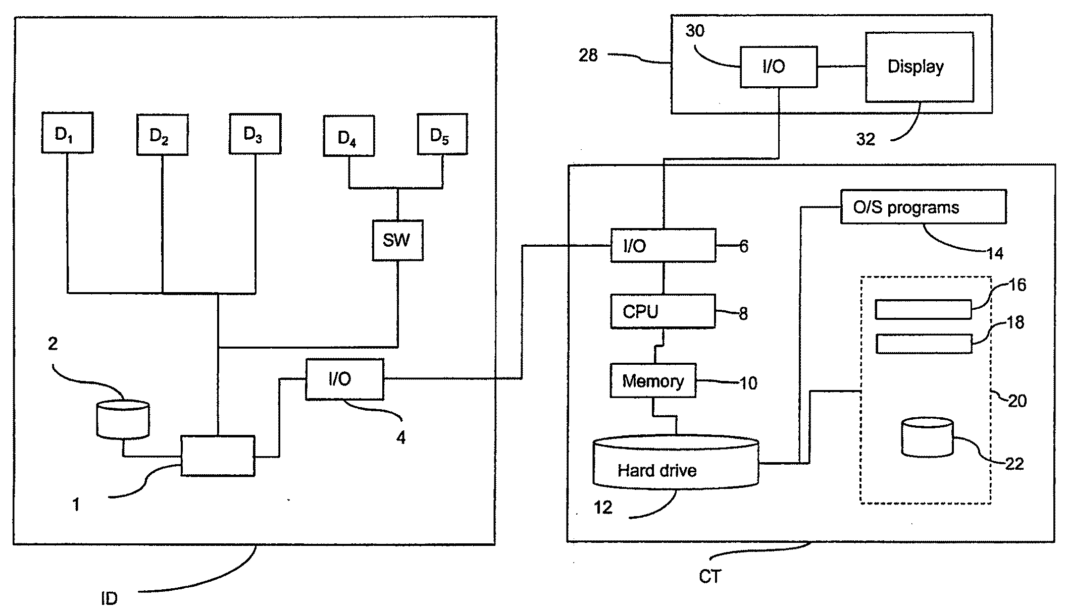
[0061]The imaging system will be described with reference to FIG. 1 which shows a schematic illustration of data processing components of an imaging system arranged in accordance with the invention. The system includes an imaging device ID, such as a confocal microscope, as described in further detail below, which includes detectors D1, D2, D3, D4, D5, a switch SW, a control unit 1, an image data store 2 and an Input / Output (I / O) device 4. An associated computer terminal CT includes a central processing...


PUM

PropertyMeasurementUnit
areasaaaaaaaaaa
adhesiveaaaaaaaaaa
fluorescentaaaaaaaaaa

Abstract

A method of imaging one or more biological objects using imaging apparatus capable of capturing an image across an imaging area. The method includes: placing the one or more biological objects (236) in an environment; providing in the environment, outside of the one or more biological objects, a contrast enhancing agent which provides contrast in an image between the one or more biological objects and the environment; and recording an image (240) of the one or more biological objects and the environment using the imaging apparatus, whereby a spatial definition for said one or more biological objects is derivable using contrast in the image which is provided by the contrast enhancing agent.

Description

FIELD OF THE INVENTION[0001]The invention relates to a method of imaging one or more biological objects using imaging apparatus. The invention further relates to computer software and apparatus arranged to perform such a method.BACKGROUND OF THE INVENTION[0002]There is currently a need in drug discovery and development and in general biological research for methods and apparatus for accurately performing cell-based assays. Cell-based assays are advantageously employed for assessing the biological activity of chemical compounds.[0003]In addition, there is a need to quickly and inexpensively screen large numbers of chemical compounds. This need has arisen in the pharmaceutical industry where it is common to test chemical compounds for activity against a variety of biochemical targets, for example, receptors, enzymes and nucleic acids. These chemical compounds are collected in large libraries, sometimes exceeding one million distinct compounds.[0004]Performing cell-based assays often i...

Claims


Application Information

Patent Timeline
Patent Type & Authority Applications(United States)
IPC IPC(8): G06K9/00G01N15/14G01N21/03G01N21/64G06T5/00
CPCG01N15/1475G01N21/6452G01N21/6456G01N21/6486G06T2207/30024G06T7/0081G06T2207/10056G06T2207/10064G06K9/00127G06T7/11G06V20/69
Inventor ADIE, ELAINE J.ARINI, NICHOLAS S.
Owner GE HEALTHCARE LTD
Why Eureka
Try Eureka