
Medical device for insertion into a joint

a medical device and joint technology, applied in the direction of shoulder joints, joint implants, prostheses, etc., can solve the problems of affecting the quality of life of patients, so as to reduce the number of particles produced.

Inactive Publication Date: 2009-09-17
DSM IP ASSETS BV
View PDF5 Cites 45 Cited by
  • Summary
  • Abstract
  • Description
  • Claims
  • Application Information

AI Technical Summary

Benefits of technology

The present invention relates to medical devices that can be used in damaged joints involving articulating surfaces. The device is made up of a fabric or network of polymer fibers and / or metal fibers that are used to fill the volume within the device. The device can be made from a single material or a combination of materials. The device can be coated or covered with a polymer material that optionally includes metal components. The device can have a smooth surface and can be equipped with an inner volume that contains a network of polymer fibers and / or metal fibers. The device can also have a self-healing surface that can be triggered by injuries. The invention also includes two-part or unit medical devices that fit into each other.

Problems solved by technology

However, serious complications are caused by the replacement of artificial joints, in particular a high occurrence rate of loosening problems resulting in breakage of the bones around the artificial joint.
In particular, the invasive character of the fixation of the prostheses such as anchoring of the prosthesis with screws and pins results in numerous side-effects such as risk of infection, loosening as mentioned above, damage on excising bone due to interruption of blood supply and necrosis.
Weight-bearing joints, in which movement in more than one direction takes place, are normally rather difficult to replace.
Improving wear resistance without losing strength or causing oxidative degradation is a difficult balance to obtain.
These particles can give rise to inflammation within the individual where the implant is implanted.
Still the produced particles give rise to inflammation and also there is a risk that the implant becomes loose.

Method used


Image

  • Medical device for insertion into a joint
  • Medical device for insertion into a joint
  • Medical device for insertion into a joint

Examples


example 1

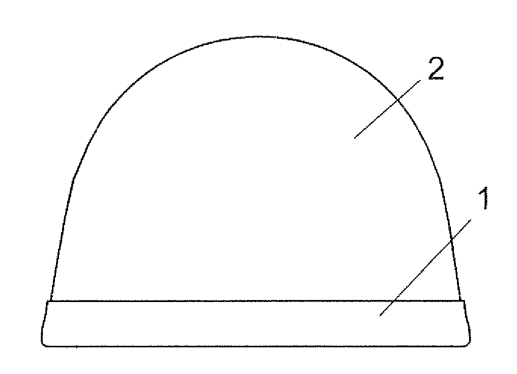
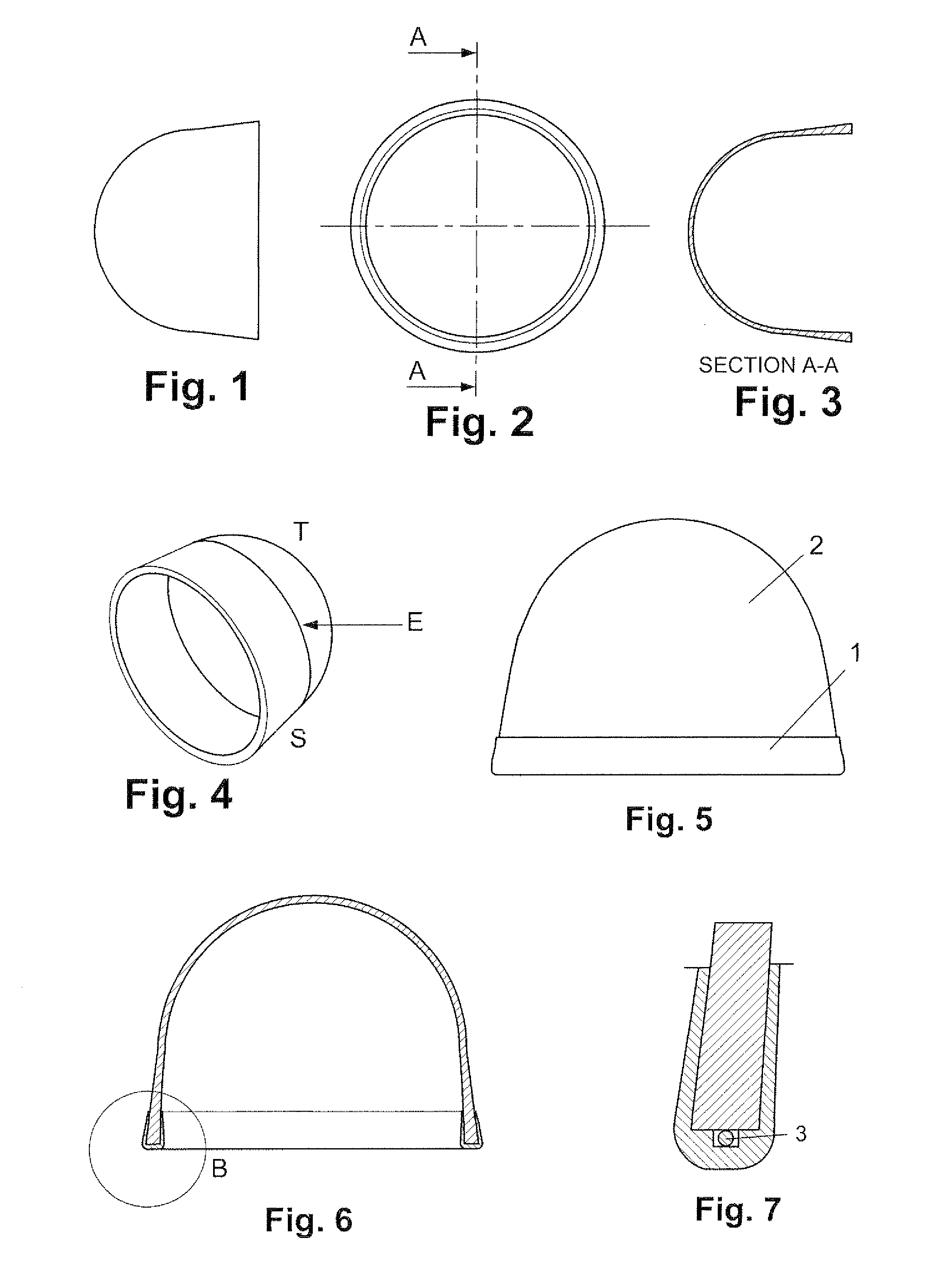
Artificial Cartilage Cup

[0678]The artificial cartilage cup is an artificial joint spacer made to replace the missing or damaged cartilage so the joint can stay mobile.

[0679]The cup is based on a sandwich construction with a LDPE core reinforced on both sides with fibre fabric.

[0680]At the edge metal markers makes it possible to trace the cup when implanted.

[0681]The round LDPE collar of the cup makes a cup without sharp edges and captures the metal markers.

[0682]Finally a crosslinking of the polymer improves the performance of the LDPE core.

[0683]The production process may include the following steps:

[0684]Injection Moulding of Base LDPE Disk

[0685]The LDPE disk is made of pellets / granulates in the injection moulding process. The disk is approximately five mm thick and 134 min in diameter. One standard disk size will later be formed to different size of cups.

[0686]Pressure Consolidation with Fibre Fabric

[0687]Two pieces of 20×20 cm fibre fabric are placed on each side of the disk and...


PUM


Abstract

The present invention relates to implants used for alleviating and / or preventing conditions relating to damaged joints involving articulating surfaces. The implants comprise fibre of polymer and / or metal, and can be used as an artificial joint, as part of an artificial joint or as an artificial joint spacer made to replace the missing cartilage or to improve the slidability between two natural and / or artificial components of the body, or between a natural and artificial component. The product of the invention can be used to partly or entirely coat medical products or to make up implant partly or entirely.

Description

CROSS REFERENCE TO RELATED APPLICATIONS[0001]This application is a continuation of application Ser. No. 11 / 917,502 that was submitted to the United States Patent and Trademark Office on Dec. 13, 2007, which is a U.S. National Stage entry under 35 U.S.C. § 371 of and claims priority to International Patent Application No. PCT / DK2006 / 000343, filed Jun. 14, 2006, designating the United States of America and published in English on Dec. 21, 2006 as WO 2006 / 133711, which claims the benefit of Denmark Application No.: PA 2005 00870, filed Jun. 14, 2005, and Denmark Application No.: PA 2005 01228, filed Sep. 2, 2005, each of which is hereby incorporated by reference in its entirety.FIELD OF INVENTION[0002]The present invention relates to a method and a medical device for alleviating and / or preventing conditions relating to damaged joints involving articulating surfaces. Especially the present invention relates to medical products which are coated partly or entirely by one or more polymeric...

Claims


Application Information

Patent Timeline
Patent Type & Authority Applications(United States)
IPC IPC(8): A61F2/30
CPCA61F2/0077A61L27/446A61F2/24A61F2/30721A61F2/30756A61F2/30767A61F2/3094A61F2/30965A61F2/32A61F2/34A61F2/38A61F2/3804A61F2/40A61F2/4202A61F2/4225A61F2/4241A61F2/4261A61F2/44A61F2/82A61F2002/30004A61F2002/30016A61F2002/3008A61F2002/30217A61F2002/30247A61F2002/3025A61F2002/30324A61F2002/30604A61F2002/30616A61F2002/307A61F2002/30701A61F2002/30703A61F2002/30705A61F2002/30713A61F2002/30754A61F2002/30757A61F2002/30957A61F2002/30971A61F2002/4253A61F2230/0067A61F2230/0071A61F2240/001A61F2240/004A61F2250/0014A61F2250/0019A61F2250/0036A61F2250/0081A61F2250/0087A61F2250/0098A61F2/12A61F2002/30242A61F2002/30245A61F2002/30593A61F2002/3071
Inventor SPORRING, SUNE LUNDOLSEN, OLELAURITZEN, JES BRUUNANDERSEN, TOM LOGSTRUPBRONDSTED, POVLBECHGAARD, KLAUSHANSEN, JAN GULDBERGJENSEN, HENRIKALMDAL, KRISTOFFERMOURITSEN, SOREN
Owner DSM IP ASSETS BV
Learn More