Medical imaging method and apparatus allowing localized image quality specification

a medical imaging and quality specification technology, applied in the field of medical imaging, can solve the problems that the dose applied in such radiography of patients with electromagnetic radiation is consistently the subject of intensive and critical discussions, and achieve the effect of simplifying the operability and clarity for the user

Active Publication Date: 2009-09-24
SIEMENS HEALTHCARE GMBH
View PDF19 Cites 23 Cited by
  • Summary
  • Abstract
  • Description
  • Claims
  • Application Information

AI Technical Summary

Benefits of technology

[0029]In an alternative embodiment, a normalized input parameter is imported. In particular a normalized input parameter is predetermined by such a normalized input parameter, which normalized input parameter is evaluated (for example using measurements on a measurement phantom) and is frequently referred to as an “average patient”. Using the normalized input parameter, it is not necessary for a physician to explicitly take into account the size and weight of a patient to be examined, for example, into account in the selection of the input parameter. Rather, in this embodiment it is possible for the physician simply to select the normalized input parameter, f

Problems solved by technology

The dose applied in such radiography of the patient with electromagnetic radia

Method used


Image

  • Medical imaging method and apparatus allowing localized image quality specification
  • Medical imaging method and apparatus allowing localized image quality specification

Examples


Embodiment Construction

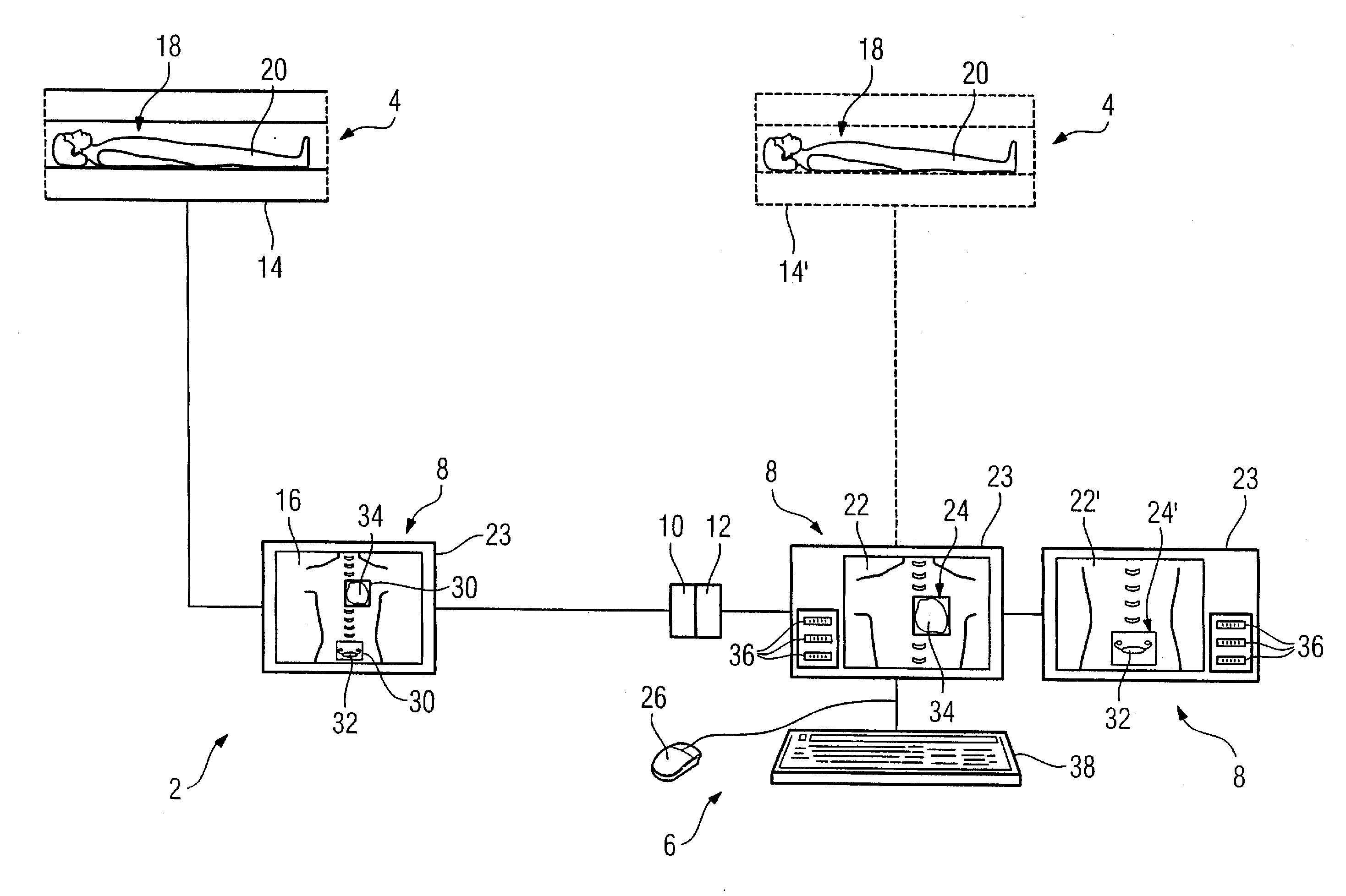
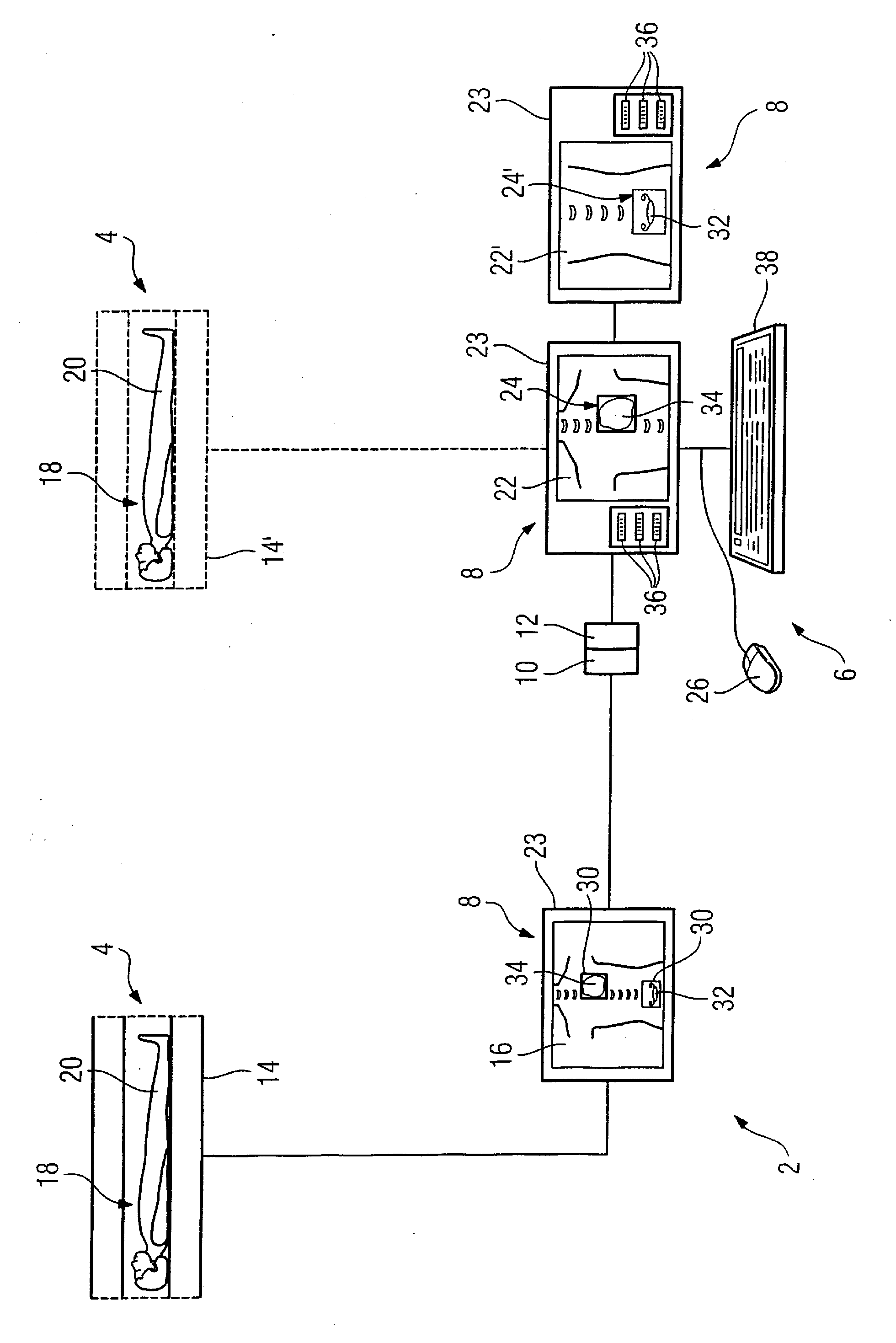
[0043]The method workflow of an embodiment of the method according to the invention will be explained using the illustration of the medical imaging device 2 in the FIGURE.

[0044]The imaging device 2 has an image acquisition apparatus 4, an importation device 6, a control device 10, and a memory element 12.

[0045]The image acquisition apparatus 4 is a computed tomography apparatus 14, called a CT apparatus 14 in the following. The CT apparatus 14 has an x-ray source and a detector (not shown), in particular in the form of a detector ring. The x-ray source and the detector are arranged opposite one another. The CT apparatus 14 is configured to implement an image acquisition 16 of an examination region 18 of a patient 20 as well as to obtain a number of planning image exposures (images) 22, 22′. Both the image acquisition 16 and the planning images 22, 22′ are visualized here on the presentation device 8 executed as a computer monitor 23.

[0046]The image exposure 16 is a three-dimensional...


PUM


Abstract

In a method and device for medical imaging, a number of input parameters with regard to an image exposure are imported into a controller of the imaging device, that associate a desired image quality with a defined image exposure region. A number of control parameters are determined corresponding to each input parameter. The controller supplies each control parameter to the image acquisition apparatus for acquiring the image exposure of the examination region with the desired local image quality.

Description

BACKGROUND OF THE INVENTION[0001]1. Field of the Invention[0002]The invention concerns a method for medical imaging as well as a corresponding medical imaging device.[0003]2. Description of the Prior Art[0004]Medical imaging is used for imaging, for implementing radiological examinations. In a radiological examination a subject (in particular a patient) is exposed with radiation. Electromagnetic beams or particle beams, for example x-rays or electrons, can be used for the exposure. As used herein, “an exposure” means radiograph of the patient.[0005]Predominantly due to different local material properties of the tissue structures located in the beam path, the incident radiation is affected to different degrees in its passage through the body of the patient. In particular, different tissue structures have different radiation attenuation properties. This causes the beams passing through the body of the patient to be attenuated to different degrees. A detector signal corresponding to th...

Claims


Application Information

Patent Timeline
IPC IPC(8): H05G1/60
CPCA61B5/055A61B5/417A61B6/032A61B6/4441A61B6/463A61B6/545A61B6/469A61B6/488A61B6/503A61B6/542A61B6/466
Inventor HARAS, GABRIEL
Owner SIEMENS HEALTHCARE GMBH
Why Eureka
Try Eureka