Apparatus and Method for Evaluating Ex Vivo Tissue Samples by Electrical Impedance

a tissue sample and electrical impedance technology, applied in the field of instruments for evaluating tissue samples in medical pathology, can solve problems such as requiring considerable skill, and achieve the effect of rapid assessment of the electrical impedance spectrum

Active Publication Date: 2009-10-08
WISCONSIN ALUMNI RES FOUND
View PDF0 Cites 25 Cited by
  • Summary
  • Abstract
  • Description
  • Claims
  • Application Information

AI Technical Summary

Benefits of technology

[0006]The present invention provides a system that can rapidly assess the electrical impedance spectrum (complex impedance as a function of frequency) of thin but large area tissue samples without sectioning or other preparation. There is currently evidence that impedance characteristics of tissue may provide a method of rapidly distinguishing benign from cancerous tumors.

Problems solved by technology

This process can be accomplished in less than an hour, but requires considerable skill.

Method used


Image

  • Apparatus and Method for Evaluating Ex Vivo Tissue Samples by Electrical Impedance
  • Apparatus and Method for Evaluating Ex Vivo Tissue Samples by Electrical Impedance
  • Apparatus and Method for Evaluating Ex Vivo Tissue Samples by Electrical Impedance

Examples


Embodiment Construction

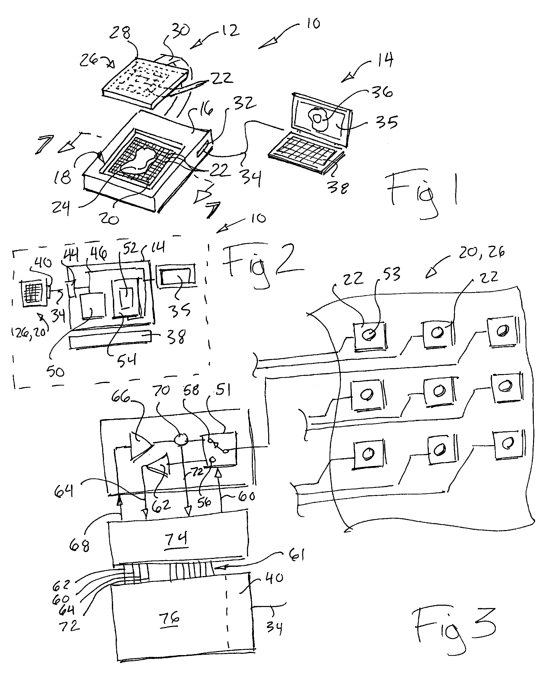
[0048]Referring now to FIG. 1, the impedance measuring apparatus 10 of the present invention may employ a tissue sample unit 12 and associated computer 14, the latter providing display and program input capabilities as will be described below. In alternative embodiments, it will be understood that the computer 14 functions may be incorporated into the tissue sample unit 12.

[0049]In the embodiment shown, the tissue sample unit 12 includes a base portion 16 having a well 81 exposing at its bottom a first planar electrode array 20 comprised of perpendicular and rectilinear rows and columns of electrodes 22 electrically isolated from each other by intervening channels. The well 18 is sized to receive an unprocessed tissue sample 24 typically several millimeters thick and no more than 1 cm thick (measured perpendicularly to the surface of the array 20) and having a height and width (measured along the surface of the array 20) of less than approximately 4 cm. The electrode array 20 is siz...


PUM

PropertyMeasurementUnit
widthaaaaaaaaaa
thickaaaaaaaaaa
frequencyaaaaaaaaaa

Abstract

A device for characterizing ex vivo tissue employs a set of independent electrodes that may be used to scan the tissue by moving a voltage gradient across the tissue surface acquiring impedance spectrographs that may be mapped to an image.

Description

CROSS REFERENCE TO RELATED APPLICATION[0001]This application claims the benefit of U.S. provisional application 61 / 072,745 filed Apr. 2, 2008 and hereby incorporated by reference.BACKGROUND OF THE INVENTION[0002]The present invention relates generally to instruments for evaluating tissue samples in medical pathology, and in particular to a device that characterizes tissue samples using precise measurements of electrical impedance of the tissue.[0003]The diagnosis of cancer and other diseases is often made by the examination of tissue samples removed from the patient during a biopsy or surgical procedure. The tissue sample may be preserved chemically and then stained and sliced into layers that are on an order of one to several cells in thicknesses. These sections are examined by a pathologist who may study these sections under a microscope to reach a conclusion about whether the tissue is cancerous.[0004]The above process may take substantial time to complete and therefore an altern...

Claims


Application Information

Patent Timeline
Patent Type & Authority Applications(United States)
IPC IPC(8): C12M1/34
CPCY10T436/2575G01N33/4836
Inventor GREGORY, WILLIAM D.
Owner WISCONSIN ALUMNI RES FOUND
Why Eureka
Try Eureka