Biopsy Devices

a biopsy device and device technology, applied in medical science, surgery, vaccination/ovulation diagnostics, etc., can solve problems such as cancerous lesions standing out under bsgi imaging

Inactive Publication Date: 2009-10-29
DEVICOR MEDICAL PROD
View PDF44 Cites 55 Cited by
  • Summary
  • Abstract
  • Description
  • Claims
  • Application Information

AI Technical Summary

Benefits of technology

[0007]The use of a biopsy device with PEM and / or BSGI may warrant features or techniques that are different from those used with other imaging modalities. For instance, with X-ray it may be desirable to have a radiopaque biopsy needle to be able to determine if the needle is in the correct location in the target tissue. To target in ultrasound, it may be desirable for a biopsy probe needle has to have a good amount of echogenecity to be visible in the modality. With MRI, the ability to see the biopsy needle in the breast may mean that there should be no artifact in the needle to affect the targeted tissue area.

Problems solved by technology

Cancerous cells may have a higher tendency to absorb certain gamma emitting radiotracers, which may result in cancerous lesions standing out under BSGI imaging.

Method used


Image

  • Biopsy Devices
  • Biopsy Devices
  • Biopsy Devices

Examples


Embodiment Construction

[0030]The following description of certain examples of the invention should not be used to limit the scope of the present invention. Other examples, features, aspects, embodiments, and advantages of the invention will become apparent to those skilled in the art from the following description, which is by way of illustration, one of the best modes contemplated for carrying out the invention. As will be realized, the invention is capable of other different and obvious aspects, all without departing from the invention. Accordingly, the drawings and descriptions should be regarded as illustrative in nature and not restrictive.

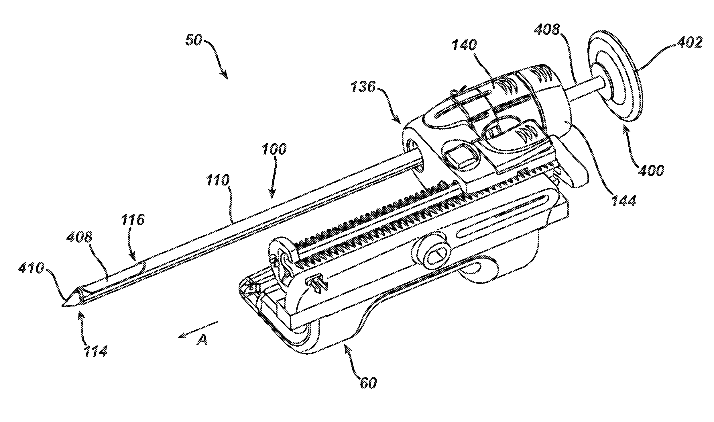
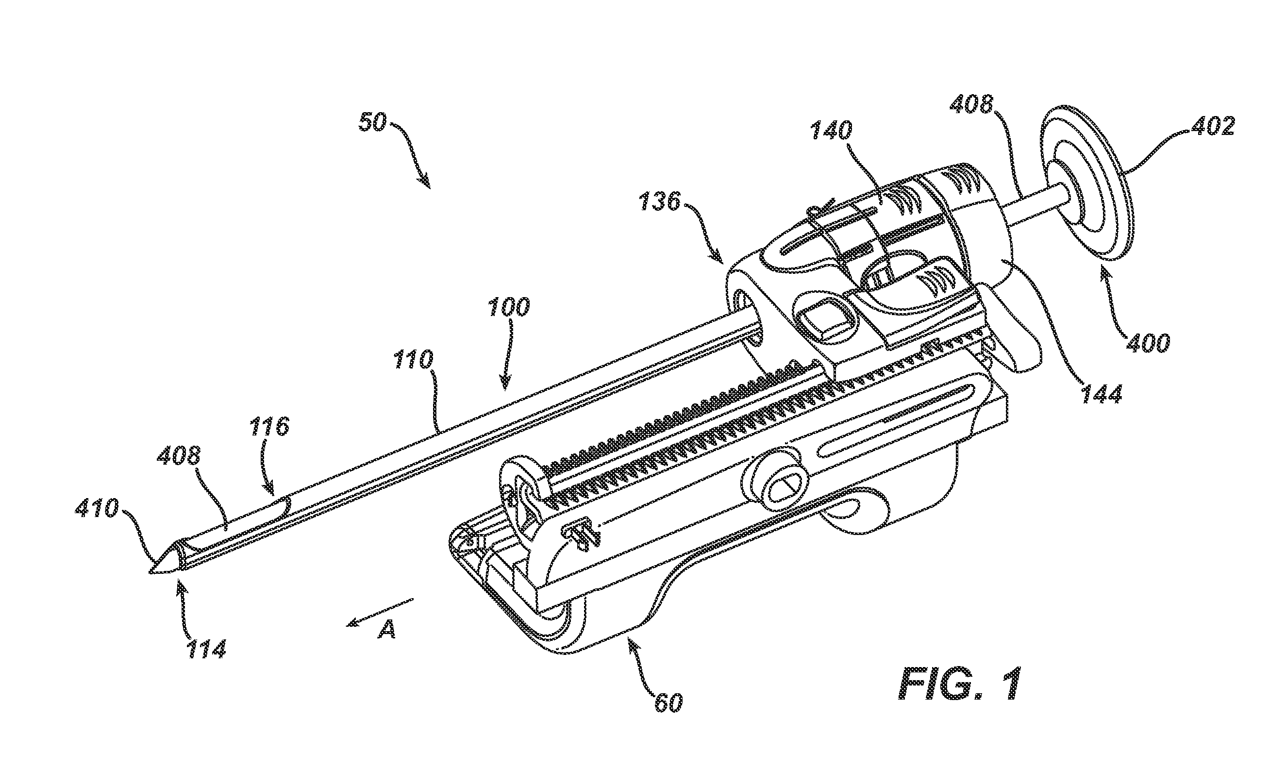
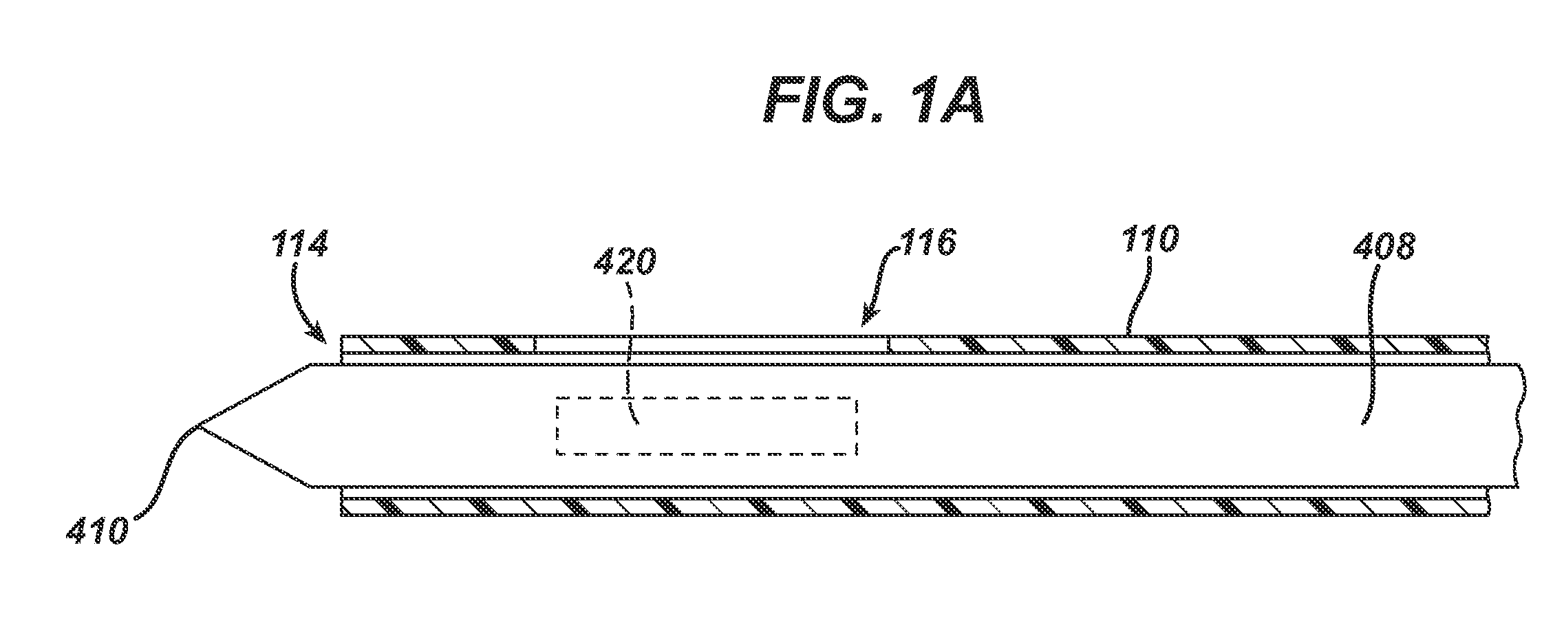
[0031]FIGS. 1 and 1A depict one biopsy targeting assembly 50 in accordance with the present invention that may be used with PEM, PET, BSGI, or other nuclear imaging systems utilizing an isotope or other radiation emitting source. The assembly shown can include similar structures employed in a targeting assembly described in one of published U.S. patent applications...


PUM


Abstract

Biopsy devices and methods useful with Positron Emission Tomography (PET) and Breast Specific Gamma Imaging (BSGI) are disclosed. A biopsy device including a flexible tube having a side aperture, and a PET or BSGI imageable material disposed within the flexible tube is disclosed. A biopsy method is disclosed that includes advancing a flexible tube having a PET or BSGI imageable material distally through the biopsy device. Various other embodiments and applications are disclosed.

Description

[0001]This application claims priority to U.S. Provisional Application 61 / 047,160 filed Apr. 23, 2008.BACKGROUND[0002]Biopsy samples have been obtained in a variety of ways using various devices. An exemplary biopsy device is the MAMMOTOME device from Ethicon Endo-Surgery, Inc. of Cincinnati, Ohio. Further exemplary biopsy devices are disclosed in U.S. Pat. No. 5,526,822, entitled “Method and Apparatus for Automated Biopsy and Collection of Soft Tissue,” issued Jun. 18, 1996; U.S. Pat. No. 6,086,544, entitled “Control Apparatus for an Automated Surgical Biopsy Device,” issued Jul. 11, 2000; U.S. Pub. No. 2003 / 0109803, entitled “MRI Compatible Surgical Biopsy Device,” published Jun. 12, 2003; U.S. Pub. No. 2007 / 0118048, entitled “Remote Thumbwheel for a Surgical Biopsy Device,” published May 24, 2007; U.S. Provisional Patent Application Ser. No. 60 / 869,736, entitled “Biopsy System,” filed Dec. 13, 2006; U.S. Provisional Patent Application Ser. No. 60 / 874,792, entitled “Biopsy Sample ...

Claims


Application Information

Patent Timeline
Patent Type & Authority Applications(United States)
IPC IPC(8): A61B10/02
CPCA61B10/0275A61B10/0283A61B19/54A61B2017/3411A61B2019/462A61B6/037A61B2019/542A61B17/3403A61B2019/5454A61B2019/5404A61B2019/5408A61B2090/3904A61B2090/392A61B2090/062A61B90/39A61B2090/3908A61B2090/3954
Inventor LEIMBACH, JESSICA P.RHAD, EDWARD A.
Owner DEVICOR MEDICAL PROD
Why Eureka
Try Eureka