
Use of the chaperone receptor-associated protein (RAP) for the delivery of therapeutic compounds to the brain and other tissues

Inactive Publication Date: 2005-02-03
HORIZON ORPHAN LLC
View PDF18 Cites 117 Cited by
  • Summary
  • Abstract
  • Description
  • Claims
  • Application Information

AI Technical Summary

Benefits of technology

[0013] In one aspect, the invention provides compounds comprising RAP or a RAP polypeptide conjugated to a therapeutic and / or diagnostic / investigational agent and pharmaceutical compositions of such compounds. In some embodiments, the RAP or RAP polypeptide conjugate according to the invention may be modified as desired to enhance its stability or pharmacokinetic properties (e.g., PEGylation of the RAP moiety of the conjugate, mutagenesis of the RAP moiety of the conjugate). In some preferred embodiments, the agent is a bioactive protein or peptide covalently linked to the RAP or RAP polypeptide moiety of the compound. Such conjugates may be formed by synthetic chemical reactions or joined by linker groups. In preferred embodiments, when the active agent is a protein or enzyme, the protein or enzyme is the human enzyme, a fragment of the human protein or enzyme having a biological activity of a native protein or enzyme, or a polypeptide that has substantial amino acid sequence homology with the human protein or enzyme. In some embodiments, the agent is a protein of human or mammalian sequence, origin or derivation. In some embodiments, the compound is a fusion protein of RAP or a RAP polypeptide portion and an active agent protein or polypeptide portion. The agent polypeptide portion of the fusion protein may be a substance having therapeutic activity such as a growth factor, lymphokine or peptide drug. The agent may be an enzyme or other bioactive protein or polypeptide. In other embodiments, the agent is an enzyme or protein whose deficiency causes a human disease such as Pompe's disease (e.g. alpha-glucosidase). In other embodiments, the enzyme is selected for its beneficial effect. In other embodiments, the conjugate is formed by non-covalent bonds between the carrier and an antibody to which the active agent is attached.
[0022] In other aspects, the invention provides methods of treating tissues or organs having proportionately greater, preferably more than two-fold, amounts of LRP receptors on their cells than other tissues or organs. The selective biodistribution of RAP or RAP-polypeptide conjugated active agents can enhance the selective targeting of such conjugated agents to specific organs.
[0031] In another embodiment, the present invention provides for a method for identifying an agent that can prevent, ameliorate or treat a lysosomal storage disease, by administering RAP or a RAP polypeptide conjugated enzyme to a cell, wherein absence of the enzyme causes the lysosomal storage disease; and determining whether the agent reduces damage to the cell compared to damage to the cell if the conjugated agent was not administered to the cell. In certain embodiments, the method is a high throughput assay.

Problems solved by technology

The blood-brain barrier (BBB) also impedes access of beneficial active agents (e.g., therapeutic drugs and diagnostic agents) to central nervous system (CNS) tissues, necessitating the use of carriers for their transit.
For example, management of the neurological manifestations of lysosomal storage diseases (LSDs) is significantly impeded by the inability of therapeutic enzymes to gain access to brain cell lysosomes.
LSDs are characterized by the absence or reduced activity of specific enzymes within cellular lysosomes, resulting in the accumulation of undegraded “storage material” within the intracellular lysosome, swelling and malfunction of the lysosomes, and ultimately cellular and tissue damage.
However, the BBB blocks the free transfer of many agents from blood to brain, and LSDs that present with significant neurological sequelae (e.g. MPS III, MLD, GM1) are not expected to be as responsive to intravenous ERT.
Direct injection of an active agent into brain tissue bypasses the vasculature completely, but suffers primarily from the risk of complications (infection, tissue damage) incurred by intra-cranial injections and poor diffusion of the active agent from the site of administration.
High plasma osmolarity leads to dehydration of the capillary endothelium with partial collapse of tight junctions, little selectivity in the types of blood-borne substances that gain access to the brain under these conditions, and damage over the course of a life-long regimen of treatment.

Method used


Image

  • Use of the chaperone receptor-associated protein (RAP) for the delivery of therapeutic compounds to the brain and other tissues
  • Use of the chaperone receptor-associated protein (RAP) for the delivery of therapeutic compounds to the brain and other tissues
  • Use of the chaperone receptor-associated protein (RAP) for the delivery of therapeutic compounds to the brain and other tissues

Examples


example ii

Construction, Expression, Purification and Characterization of RAP Fusions

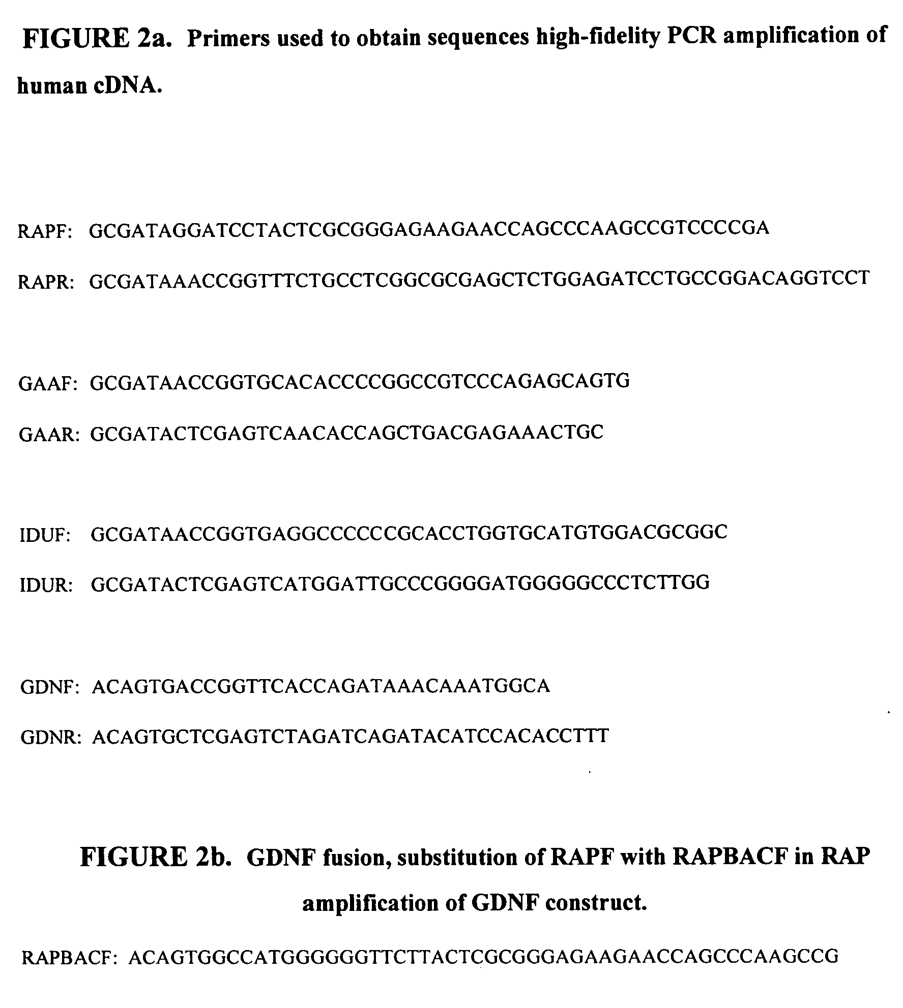
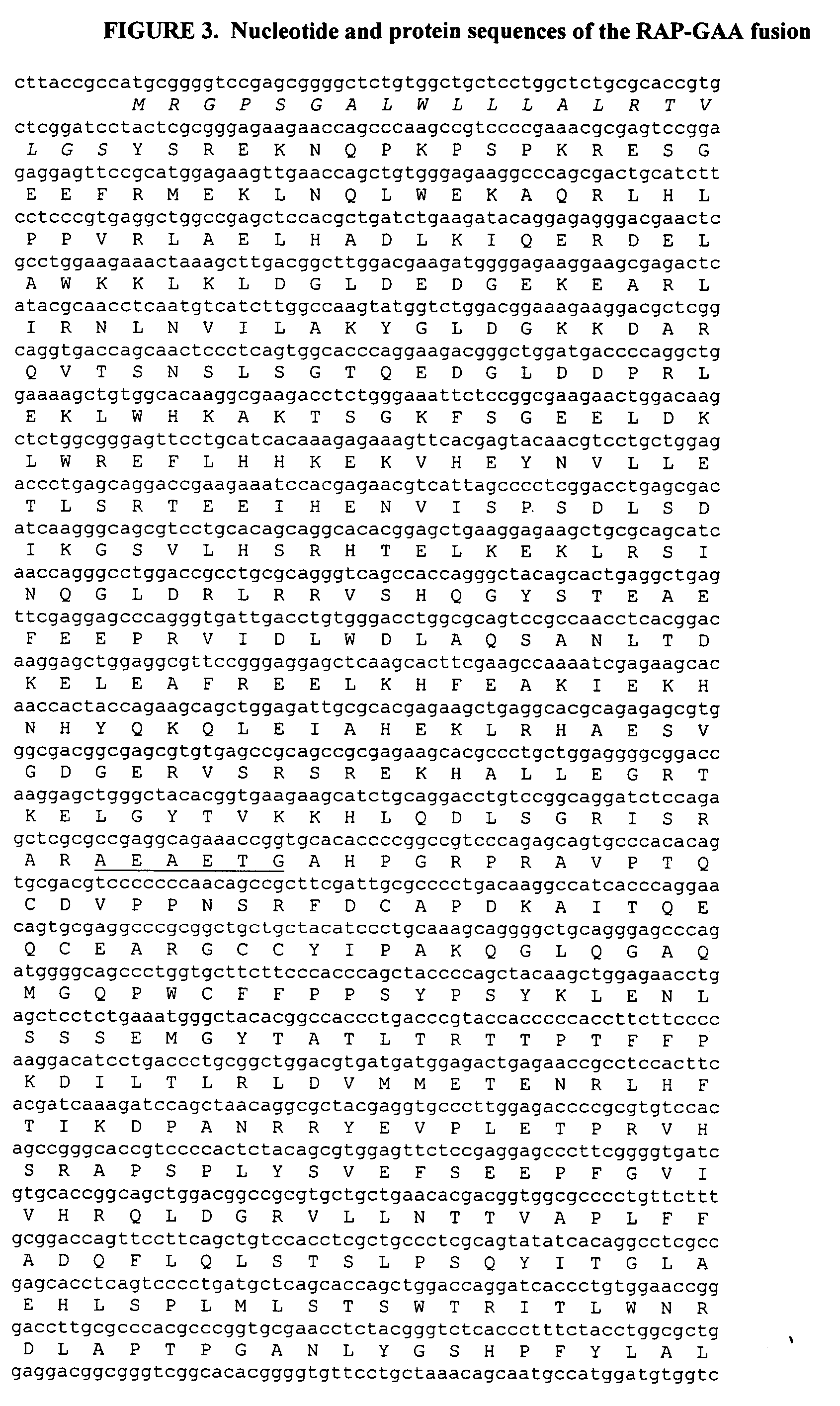
[0250] Expression constructs encoding fusions between the human receptor-associated protein (RAP) and human alpha-glucosidase (GAA), alpha-L-iduronidase (IDU) or glial cell-derived neurotrophic factor (GDNF) were prepared. For this purpose, a sequence that encodes RAP was fused to the 5′-end of sequences that encode the different fusion partners. All sequences were obtained by high-fidelity PCR amplification of human cDNA with the following primers shown in FIG. 2a. The GDNF fusion was designed for expression in bacteria. To this end, primer RAPBACF was substituted for RAPF in the RAP amplification for this construct (FIG. 2b).

[0251] The 5′-end of RAP was truncated to remove the signal peptide sequence. Instead, an in-frame BamHI site, which encodes the dipeptide GS, was added for the mammalian expression construct. Sequence encoding the tetrapeptide MGGS with an NcoI site at the 5′-end was added for the bac...

example iii

Uptake and Distribution of Unconjugated RAP to the Brain

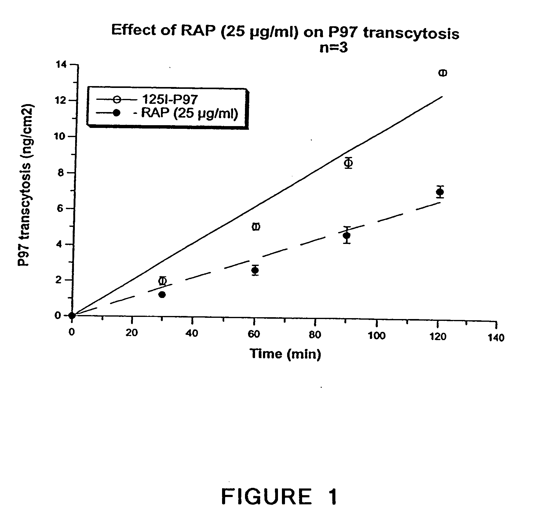
[0263] The distribution of RAP to brain was measured using a mouse in situ perfusion model. Volumes of distribution (Vd) for RAP, the positive control transferrin and the negative control albumin, were determined over a perfusion interval of 5 minutes. In addition, the relative quantities of the test proteins in the vascular and parenchymal fractions of the perfused brain were determined using the capillary depletion technique (Gutierrez, et al., J. Neuroimmunology 47(2):169-76 (1993)). The results shown in FIG. 11 include an observed, corrected Kinflux of 1 μL / g / min for transferrin. RAP had an observed, corrected Kinflux of 2.2 μL / g / min. RAP is taken up into brain.

[0264] A separate experiment was carried out at a single, 5-minute time-point to determine whether RAP is able to traverse the brain vasculature and enter the parenchyma. Brains were harvested as before, but were subjected to a capillary depletion procedure to dete...

example iv

Measurement of Specific Uptake of RAP-GAA into Enzyme-Deficient Patient Fibroblasts

[0265] The uptake of RAGA into cells deficient in GAA was characterized. The cell line used was GM244 (Coriell Cell Repository), a primary cell line isolated from a patient with glycogen storage disorder type II (Pompe's disease). These fibroblasts take up phosphorylated, recombinant GAA via the mannose-6-phosphate receptor, but also have LRPI receptors, which bind RAP. In order to identify the receptors involved in uptake of different test ligands, samples containing excess free RAP or mannose-6-phosphate were prepared.

[0266] Dilutions of RAP-GAA were made in the uptake medium (Dulbecco's Modified Eagle's Medium supplemented with 25 mM HEPES pH 7.0, 2 mM L-glutamine and 250 pg / mL bovine serum albumin) to yield fusion protein concentrations of 33, 11, 3.7, 1.2, 0.4, and 0.1 nM. The effect of 3 mM mannose-6-phosphate, 500 nM RAP and a combination of the two on the uptake of 5 nM RAP-GAA was also assa...


PUM

PropertyMeasurementUnit
Volumeaaaaaaaaaa
Timeaaaaaaaaaa
Timeaaaaaaaaaa

Abstract

This invention provides compounds of conjugates of therapeutic or active agents with RAP or a RAP polypeptide, their pharmaceutical compositions and methods for using the such compounds and compositions in the diagnosis, prophylaxis, or treatment of diseases and conditions, including particularly diseases of the central nervous system or lysosomal storage diseases.

Description

CROSS-REFERENCE TO RELATED APPLICATIONS [0001] This application is a continuation-in-part of U.S. patent application Ser. No. 10 / 206,448, filed on Jul. 25, 2002, which claims the benefit of U.S. Provisional Patent Application No. 60 / 308,002, filed Jul. 25, 2001. The contents of these and all other U.S. patents cited herein are each hereby incorporated by reference in their entirety.FIELD OF THE INVENTION [0002] The present invention is related to compositions comprising Receptor-Associated Protein (RAP) linked to a therapeutic and / or diagnostic / investigational agent, and methods of using such compounds. BACKGROUND OF THE INVENTION [0003] The brain is shielded against potentially harmful substances by the blood-brain barrier (BBB). The microvascular barrier between blood and brain is made up of a capillary endothelial layer surrounded by a basement membrane and tightly associated accessory cells (pericytes, astrocytes). The brain capillary endothelium is much less permeable to low-mo...

Claims


Application Information

Patent Timeline
IPC IPC(8): A61K38/17A61K39/395A61K48/00C07K14/705
CPCA61K38/00A61K47/48246C07K2319/23C07K2319/00C07K2319/22C07K14/705A61K47/64A61P25/00A61P25/02A61P25/16A61P25/18A61P25/28A61P35/00A61P9/10
Inventor ZANKEL, TODDSTARR, CHRISTOPHERGABATHULER, REINHARD
Owner HORIZON ORPHAN LLC
Learn More