Biopsy Marker with In Situ-Generated Imaging Properties

Active Publication Date: 2010-01-14
CR BARD INC
View PDF102 Cites 32 Cited by
  • Summary
  • Abstract
  • Description
  • Claims
  • Application Information

AI Technical Summary

Benefits of technology

[0018]A biopsy marker, preferably a breast biopsy marker, has radio-opaque properties that are derived in situ, preferably based on a natural a biological response, such as calcification or accumulation or tissue-concentration of a chemical agent that acts as an imaging contrast. In an embodiment, a biodegradable foam such as collagen foam or gelatin foam is embedded with a biological tissue that is susceptible to the calcification. The biopsy marker is

Problems solved by technology

It is believed that elastin rich tissue

Method used


Image

  • Biopsy Marker with In Situ-Generated Imaging Properties
  • Biopsy Marker with In Situ-Generated Imaging Properties

Examples


Embodiment Construction

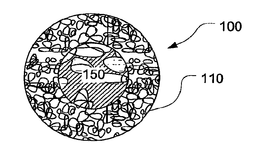
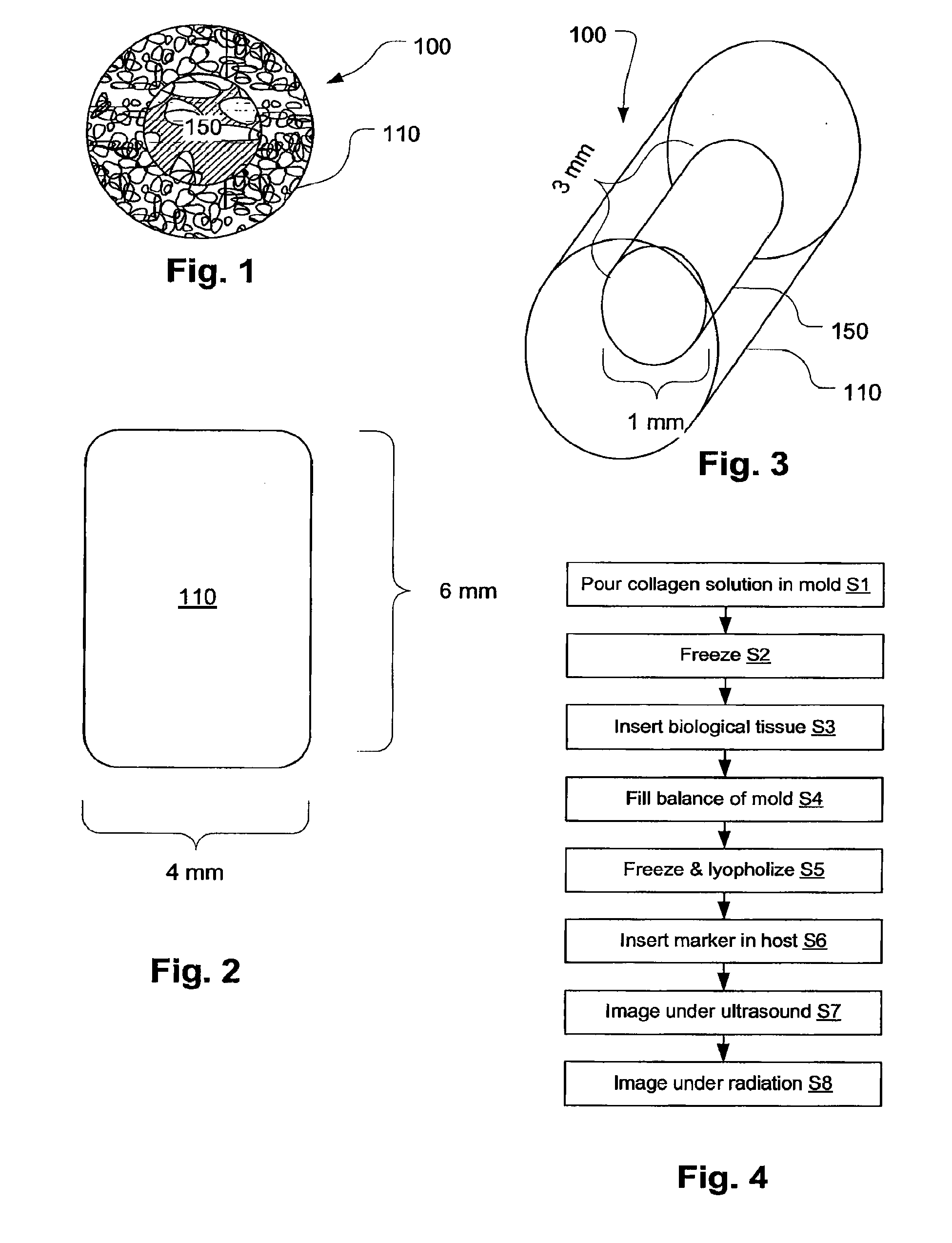
[0031]A biopsy marker, preferably a breast biopsy marker, has radio-opaque properties that are derived in situ, preferably based on a natural a biological response, such as calcification or accumulation or tissue-concentration of a chemical agent that acts as an imaging contrast. In an embodiment, a biodegradable foam such as collagen foam or gelatin foam is embedded with a biological tissue that is susceptible to the calcification. The biopsy marker is implanted to mark the biopsy site. The foam material provides ultrasonic visibility to access the implantation site. The biological tissue undergoes calcification in 30 days to 5 years depending on the chemistry of biological tissue used. The calcification generated in the biological tissue provides visibility in magnetic resonance imaging (MRI) and X-ray imaging. As a result, the marker may be located using radiation-based imaging or ultrasonic imaging.

[0032]Many types of implantable tissues can be used to prepare a biopsy marker de...


PUM

PropertyMeasurementUnit
Timeaaaaaaaaaa
Shapeaaaaaaaaaa
Biological propertiesaaaaaaaaaa

Abstract

A biopsy marker having radio-opaque properties that are derived in situ, based on a natural a biological response, such as for example, calcification, accumulation or tissue-concentration of a chemical agent so as to provide an imaging contrast. A biodegradable foam such as collagen foam or gelatin foam is embedded with a biological tissue that is susceptible to the calcification. Initially the marker can be imaged using ultrasound, but over time, the embedded material calcifies causing it to become visible under radiation imaging.

Description

PRIORITY DATA AND INCORPORATION BY REFERENCE[0001]This application is a U.S. nation phase of International Application No. PCT / US2007 / 087768, filed Dec. 17, 2007, which claims priority to U.S. Provisional Patent Application No. 60 / 870,502, filed Dec. 18, 2006, which are incorporated by reference in their entirety.TECHNICAL FIELD[0002]The invention relates generally to a biopsy tissue markers. More specifically, the invention further relates to a biocompatible tissue site marker that is visible under various modes of imaging.BACKGROUND[0003]Advances in modem medical imaging technologies such as X-ray, ultrasound, or magnetic resonance imaging make it possible to identify and to biopsy tumors while they are still small. When dealing with a small tumor, especially after a portion of the tumor has been removed for biopsy, it is sometimes difficult to relocate the tumor at a later time for treatment. This is particularly true in the case of tumors in the breast, where the ability to visu...

Claims


Application Information

Patent Timeline
IPC IPC(8): A61B6/00
CPCA61B19/54A61B2019/5466A61B2019/5425A61B2019/5495A61F2250/0098A61K49/0002A61K49/0409A61K49/18A61K49/222A61B5/055A61B5/062A61B5/064A61B6/12A61B8/0833A61B10/02A61B2019/5408A61B2090/392A61B2090/3995A61B90/39A61B2090/3908A61B2090/3925A61B2090/3966
Inventor TALPADE, DNYANESH A.PATHAK, CHANDRASHEKHAR
Owner CR BARD INC
Why Eureka
Try Eureka