
System, device, method, computer-readable medium, and use for in vivo imaging of tissue in an anatomical structure

a tissue anatomical and imaging technology, applied in the field of medical imaging, can solve the problems of many false negatives, limited penetration depth of optical imaging technology, and presented fluorescence imaging technology, and achieve the effect of reducing the number of false negatives

Inactive Publication Date: 2010-03-04
KONINKLIJKE PHILIPS ELECTRONICS NV
View PDF9 Cites 18 Cited by
  • Summary
  • Abstract
  • Description
  • Claims
  • Application Information

AI Technical Summary

Benefits of technology

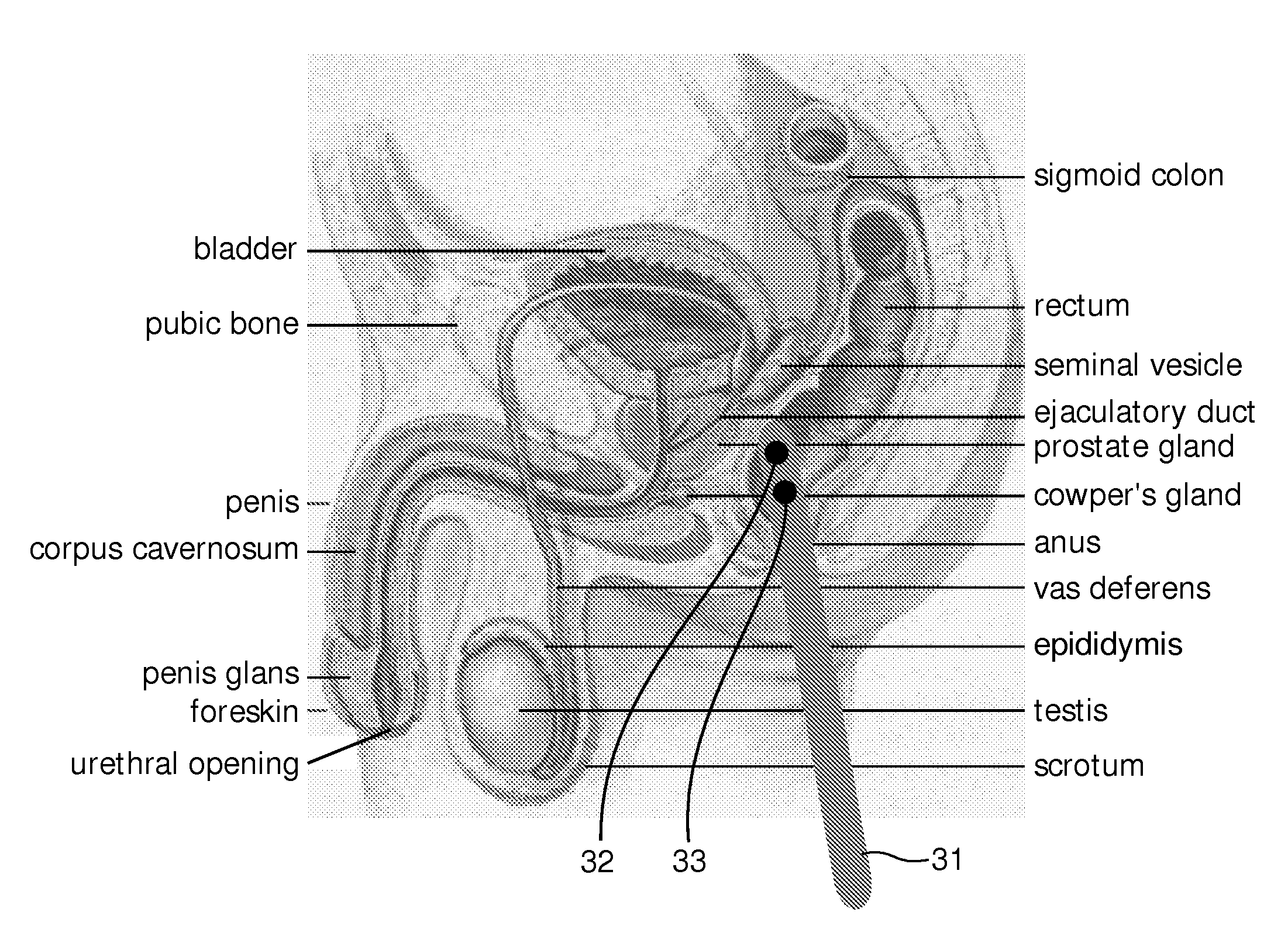
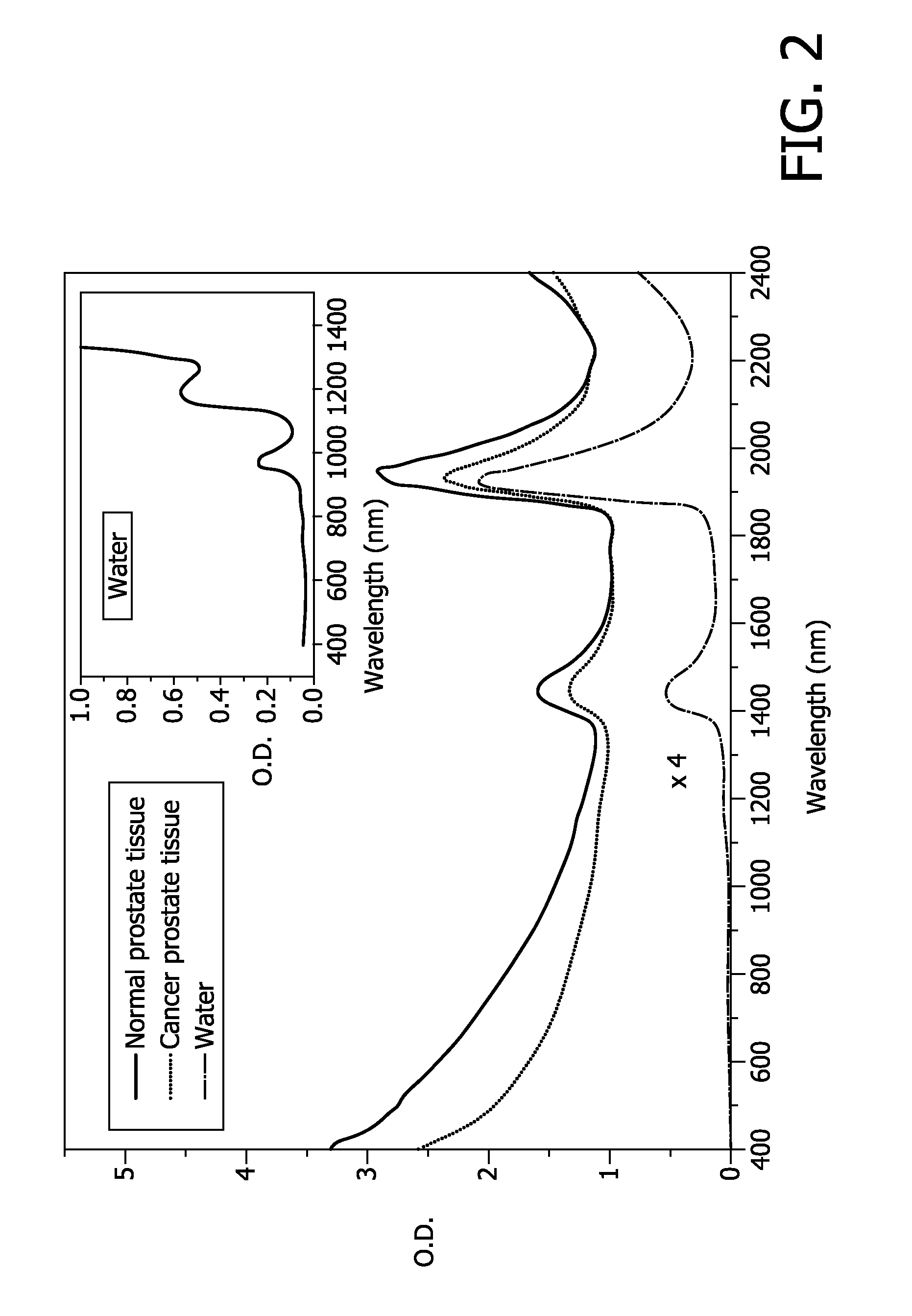
[0012]Embodiments of the present invention pertain to the use of Photoacoustic Imaging for creating an image dataset for the detection of suspicious prostate tissue. The system according to some embodiments may be used to guide a biopsy thereby reducing the number of false negatives, as the location of diseased tissue becomes known.

Problems solved by technology

However, a problem is to take a biopsy from the correct tissue volume.
Obviously, this procedure leads to numerous false negatives.
A drawback of the system is that the optical imaging technology presented, i.e. fluorescence imaging, therein only has a rather limited penetration depth.

Method used


Image

  • System, device, method, computer-readable medium, and use for in vivo imaging of tissue in an anatomical structure
  • System, device, method, computer-readable medium, and use for in vivo imaging of tissue in an anatomical structure
  • System, device, method, computer-readable medium, and use for in vivo imaging of tissue in an anatomical structure

Examples


Embodiment Construction

[0021]Several embodiments of the present invention will be described in more detail below with reference to the accompanying drawings in order for those skilled in the art to be able to carry out the invention. The invention may, however, be embodied in many different forms and should not be construed as limited to the embodiments set forth herein. Rather, these embodiments are provided so that this disclosure will be thorough and complete, and will fully convey the scope of the invention to those skilled in the art. The embodiments do not limit the invention, but the invention is only limited by the appended patent claims. Furthermore, the terminology used in the detailed description of the particular embodiments illustrated in the accompanying drawings is not intended to be limiting of the invention.

[0022]The following description focuses on embodiments of the present invention applicable to an imaging system and in particular to an imaging system for imaging of diseased tissue in...


PUM


Abstract

A system is provided that may be used for locating and diagnosing lesions in the human body in vivo. In some embodiments, once the exact position of a lesion is found, a biopsy may be taken from the lesion using e.g. ultra sound techniques for guidance of the biopsy needle. Use of the system drastically reduces the negative biopsy samples compared to currently used “blind sampling” techniques. This reduces patient discomfort and minimizes infections as the number of biopsy samples is reduced. A method and computer-readable medium is also provided.

Description

FIELD OF THE INVENTION[0001]This invention pertains in general to the field of medical imaging. More particularly the invention relates to imaging of different tissue types in vivo and guiding a tissue biopsy using medical imaging.BACKGROUND OF THE INVENTION[0002]Prostate cancer is the most common cancer in men excluding skin cancer. The American Cancer Society, ACS, estimates that about 232,090 new cases of prostate cancer will be diagnosed in the United States and 30,350 men will die of this disease in 2005. The ACS estimates that a male in the US has a 1 in 6 risk of developing prostate cancer during his lifetime.[0003]There are several tests are available for detection of prostate cancer, such as, Prostate-specific antigen (PSA) blood test, Digital Rectal Exam (DRE), Transrectal Ultrasound (TRUS) and Core Needle Biopsy. PSA, DRE and TRUS have limited sensitivity and / or specificity to the lesions and are mainly used to estimate the risk of having prostate cancer, depending on siz...

Claims


Application Information

Patent Timeline
Patent Type & Authority Applications(United States)
IPC IPC(8): A61B8/14A61B6/00
CPCA61B5/0084A61B5/0086A61B8/4416A61B8/12A61B5/0095A61B5/0035
Inventor BAKKER, LEVINUS PETERVAN BEEK, MICHAEL CORNELISVAN DER MARK, MARTINUS BERNARDUSVAN DEN HAM, RENEHENDRIKS, BERNARDUS HENDRIKUS WILHELMUSHOFFMANN, RALFVAN DER VAART, NIJS CORNELISVAN DER VOORT, MARJOLEIN
Owner KONINKLIJKE PHILIPS ELECTRONICS NV
Learn More