Method and apparatus for identifying regions of interest in a medical image

a technology of image and region, applied in image enhancement, image data processing, instruments, etc., can solve the problems of increased difficulty, adjustment of threshold, and adjustment of difficulty

Active Publication Date: 2010-04-08
SIEMENS MEDICAL SOLUTIONS USA INC
View PDF38 Cites 17 Cited by
  • Summary
  • Abstract
  • Description
  • Claims
  • Application Information

AI Technical Summary

Benefits of technology

[0040]This allows a selection of an ROI which prevents the in

Problems solved by technology

The issue in such threshold based segmentations is the adjustment of the threshold.
In some cases this adjustment is made difficult by the presence of other high uptake structures or features of the image adjacent to the region of interest.
The difficulty is more pronounced in 3D than in 2D since the user must check each slice over which the VOI is defined, since connectivity between voxels included in the object of interest may be present across voxels not in the current displayed slices.
This can be slow and laborious.
The problems with this approach are:the speed of the algorithm depends on the size of the containing region, and can be too slow for real-time feedback for larger regionsif the original containing region excluded part of the annotated region (for example on a different slice of the volume) it will need to be redrawn and the process repeatedmultiple user interactions (e.g. mouse clicks) are required (for example, to create the containing region and update the threshold value)
Although this method has a simpler user interface, it is also problematic since very permissive thresholds could potentially select mo

Method used


Image

  • Method and apparatus for identifying regions of interest in a medical image
  • Method and apparatus for identifying regions of interest in a medical image
  • Method and apparatus for identifying regions of interest in a medical image

Examples


first embodiment

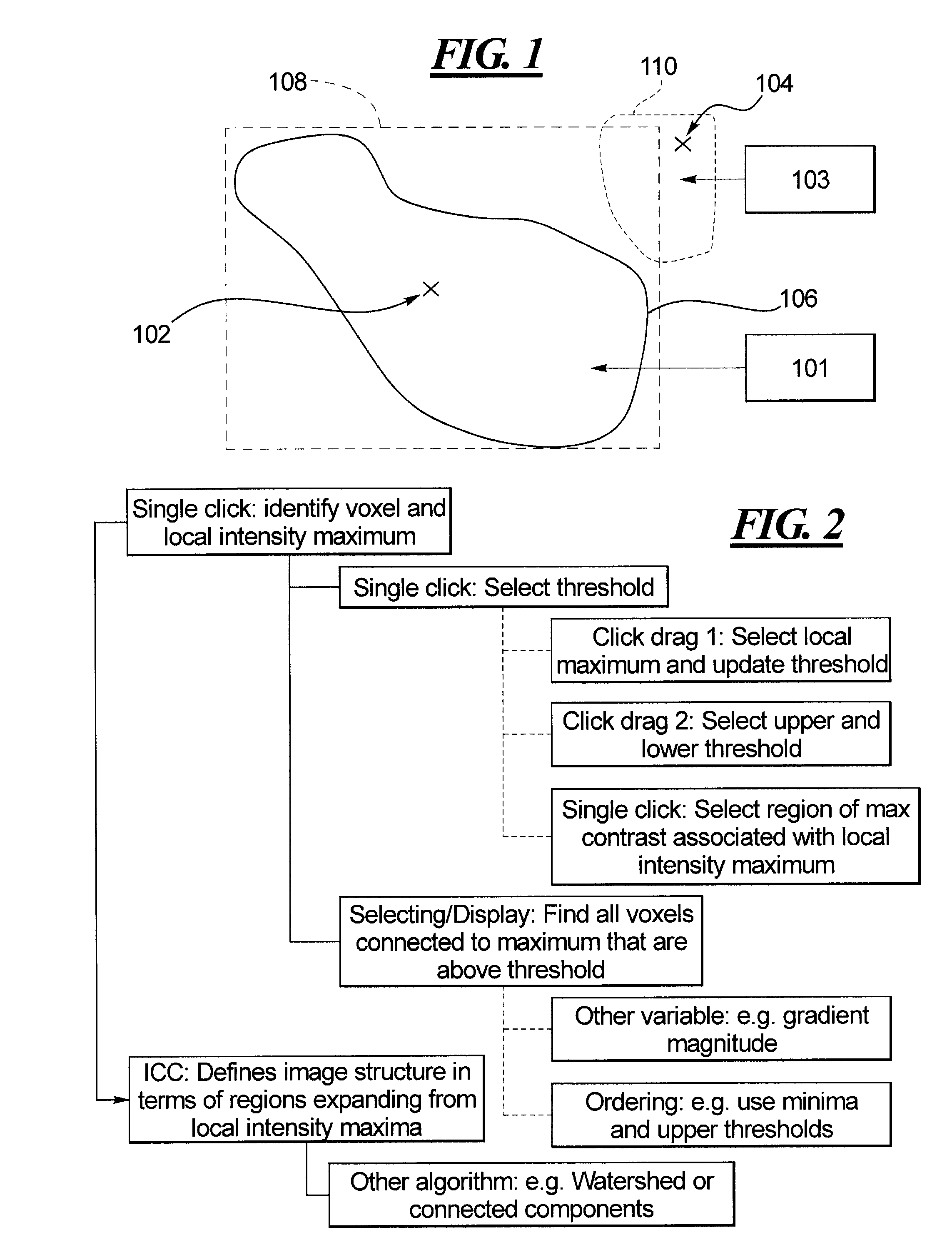
[0077]FIG. 2 sets out the processes which are generally involved in the invention. A threshold (of intensity, for example of SUV values in an FDG-PET scan) is determined from the point in the image that the user clicked on (rather than requiring a separate step to update the threshold), and the corresponding click-label is extracted from the label image, that is, which local intensity maximum is the point associated with. Next, the position of the click-point in the sorted list of voxels is found. All voxels in the intensity sorted list up until this point that correspond to the click-label are extracted. Likewise the voxels of any child-labels that have merged into the click label up until this point are extracted and stored. The collection of voxels is then highlighted.

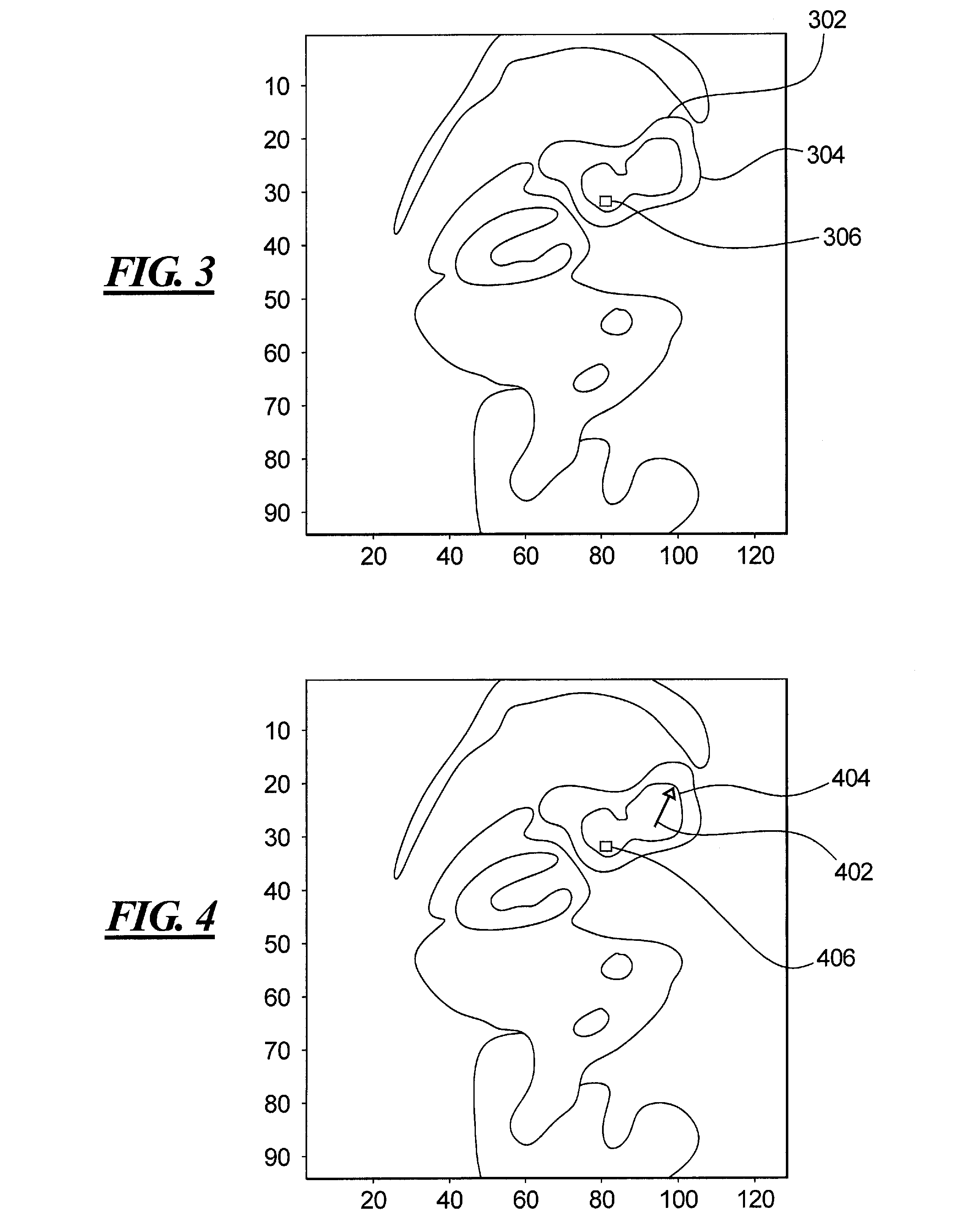
[0078]An example of the output of this process is shown in FIG. 3, which shows a coronal slice from a PET scan of a human torso. The circle 302 indicates a user click point and the black curve 304 indicates the anno...

second embodiment

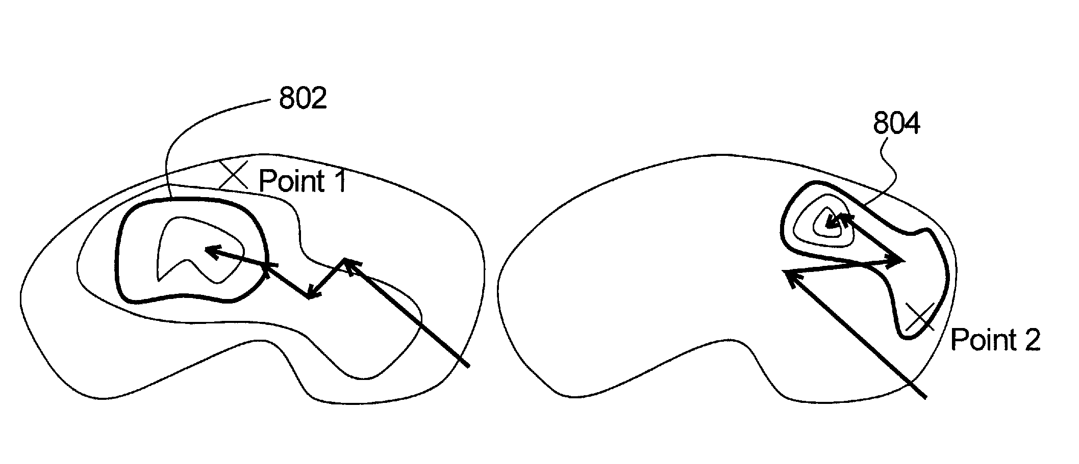
[0086]FIG. 5 sets out the processes generally involved in the present invention. The hierarchy or tree structure of the result of the ICC pre-processing is used to detect a set of candidate regions or volumes of interest (VOIs), whose relationships are encoded by the hierarchical grouping. At least two kinds of relationships can exist: one VOI can encompass another, and two VOIs can merge. These relationships are established using a variable such as intensity, or gradient magnitude.

[0087]A subset of VOIs is obtained to be presented to the user, which is also arranged as a set of tree structures. Such a subset may, for example, consist of those VOIs with the greatest contrast of intensity relative to the surrounding area, since these are deemed likely to be of interest. The user may then choose to accept some or reject others in a final group of VOIs. Alternatively, it may be possible that none of the selected subset of VOIs is sufficiently accurate in the user's judgment. In this ca...

third embodiment

[0103]FIG. 10 sets out the processes which are generally involved in the invention. This embodiment allows a freeform bounding region to be generated that delineates the largest possible region that can be selected without including additional regions, features or structures in the image which are not of interest to the user. This is referred to as the maximal bounding region. A user need not carefully select a bounding region, but rather may select any threshold (restricted automatically by the bounding region as set out below) without fear of including an extraneous feature or structure within the ROI. In addition, a connected component algorithm may not need to be re-applied if the threshold is changed.

[0104]An example of this is illustrated in 2D in FIG. 11, where the image is an isocontour representation of the image in FIG. 1. The isocontor representation is shown for convenience, since each isocontor represents a possible threshold selection from which an ROI may be obtained....


PUM


Abstract

In a method and apparatus for identifying regions of interest in medical images of a subject, in particular images captured by a medical imaging apparatus, such as a PET scanner, a list of voxels of the image is obtained, and sorted according to a first variable. A user-selection of an initial voxel in the image is then registered, and at least one voxel or group of voxels from the sorted list is selected as a region of interest, according to a property of the at least one voxel or group in relation to the user-selected initial voxel. The image may be pre-processed to generate the sorted list of voxels, following which the user selection of the initial voxel is registered.

Description

BACKGROUND OF THE INVENTION[0001]1. Field of the Invention[0002]The present invention concerns identifying regions of interest in medical images of a subject.[0003]2. Description of the Prior Art[0004]The definition of regions or volumes of interest (ROI / VOI) is a typical precursor to quantitative analysis of medical images, such as nuclear medicine emission images (for example, PET or SPECT). Such regions may be defined around areas of high intensity which correspond to high tracer uptake (hotspots). For example, in FDG-PET images for oncology, such areas may be indicative of the presence of a tumor. Oncology physicians frequently annotate lesions in PET scans for the purpose of making a diagnosis, or for use in radiotherapy. The mean or maximum tracer uptake can aid a reader in determining the likelihood of cancer. In longitudinal studies, considering the change in intensity or uptake on corresponding VOIs from images at different temporal stages may be used to determine whether a...

Claims


Application Information

Patent Timeline
IPC IPC(8): G06F3/048G06V10/25
CPCG06K9/3233G06K2009/366G06K2209/053G06T2207/30096G06T7/0091G06T2207/10104G06T2207/20101G06T7/0081G06T7/11G06T7/155G06V10/248G06V10/25G06V2201/032G06T7/0012G06V10/267
Inventor DOWSON, NICHOLAS DELANIE HIRSTWRIGHT, THOMAS GEORGEKADIR, TIMORHAKL, KEVIN SCOTT
Owner SIEMENS MEDICAL SOLUTIONS USA INC
Why Eureka
Try Eureka