

Image processing systems
a technology of image processing and image, applied in the field of medical devices, can solve the problems of affecting the fine positioning of the imaging system, cramping the working area created by such inflatable balloons, and affecting the accuracy of anatomical features in the body, so as to facilitate the physician's assessment of lesion size and facilitate tissue treatment assessment. , the effect of accurately measuring anatomical features
- Summary
- Abstract
- Description
- Claims
- Application Information
AI Technical Summary
Benefits of technology
Problems solved by technology
Method used
Image

Examples
Embodiment Construction
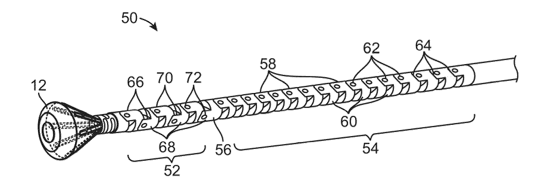
[0060]Reconfiguring a tissue visualization and treatment device from a low profile delivery configuration for intravascular delivery through the vessels of a patient to a deployed and expanded configuration may subject the distal end effector used for visualization and / or treatment, such as energy delivery, to potentially severe mechanical stresses (e.g., torsion, compression, tension, shearing, etc.). For example, a reconfigurable hood which undergoes a shape change from its collapsed configuration to an expanded conical shape may utilize a distensible, collapsible, and / or reconfigurable substrate which may utilize electrode placement and electrical connection assemblies which are robust and able to withstand such stresses. Such electrical connection assemblies may be shielded or insulated from contacting other structures so as to present a smooth or unobstructive profile for reconfiguring with the hood.
[0061]Turning now to the tissue-imaging and manipulation apparatus upon which o...
PUM
 Abstract
Description
Claims
Application Information