Method of chondrogenic differentiation from mesenchymal stem cell, and composition comprising chondrogenic cell differentiated using the method to treat disease caused by cartilage damage

Inactive Publication Date: 2010-06-03
ELECTRONICS & TELECOMM RES INST
View PDF3 Cites 1 Cited by
  • Summary
  • Abstract
  • Description
  • Claims
  • Application Information

AI Technical Summary

Benefits of technology

[0009]The present invention provides a method of chondrogenic differentiation from mesen

Problems solved by technology

However, growth factors such as TGF-β are not only expensive per se, but rather accelerate aging of cells, accompanied by a decrease in cell viability, limiting their clinical use.
Because the differentiation of human mesenchymal stem cells is more difficult than that of animal mesenchymal stem cells, there are limitat

Method used


Image

  • Method of chondrogenic differentiation from mesenchymal stem cell, and composition comprising chondrogenic cell differentiated using the method to treat disease caused by cartilage damage
  • Method of chondrogenic differentiation from mesenchymal stem cell, and composition comprising chondrogenic cell differentiated using the method to treat disease caused by cartilage damage
  • Method of chondrogenic differentiation from mesenchymal stem cell, and composition comprising chondrogenic cell differentiated using the method to treat disease caused by cartilage damage

Examples


Example

Experimental Example 1

Monolayer Culture

[0037]Human mesenchymal stem cells were purchased from LONZA (USA). The mesenchymal stem cells were attached to the bottom of a culture dish and a mesenchymal stem cell growth medium MSCGM bullet kit (LONZA, USA) was added to conduct a monolayer-culture. The culture dish was incubated in a 5% CO2 incubator at 37. The medium was exchanged once every three days, and the number of cells suitable for a three-dimensional culture was secured through a subculture performed once every week.

Example

Experimental Example 2

Three-Dimensional Culture

[0038]The mesenchymal stem cells incubated in Experimental Example 1 were treated with 0.05% trypsin-EDTA (ethylenediaminetetraacetate) and detached from the culture dish.

[0039]One group of the mesenchymal stem cells was suspended in a 2% alginate solution at a concentration of about 2×10⁶ cells / d, and cell / alginate suspensions were slowly dropped into a 102 mM CaCl2 solution and left still for 10 minutes to form spherical beads.

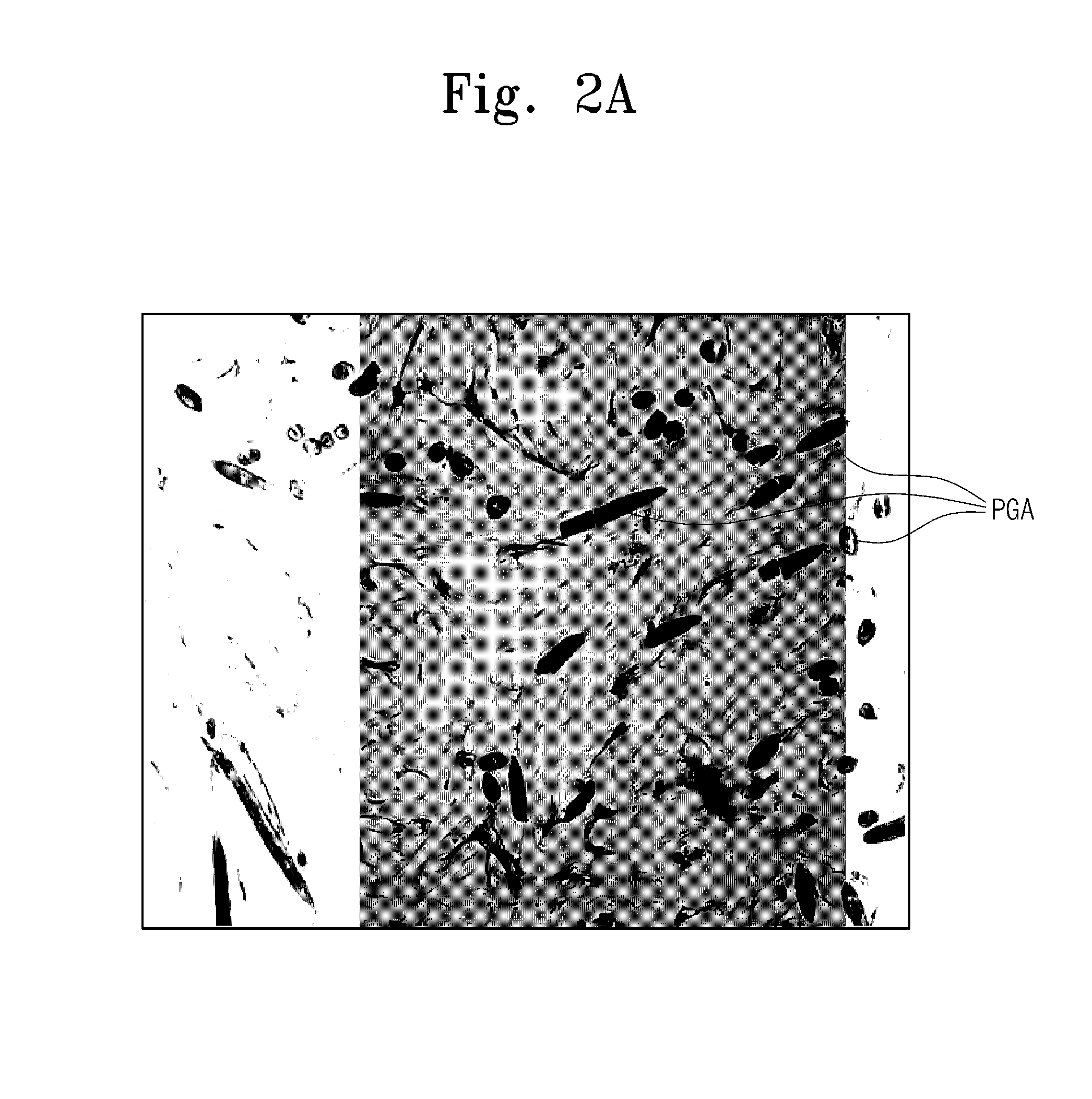
[0040]Another group of the mesenchymal stem cells, that was detached by the method for inoculation of the mesenchymal stem cells into PGA scaffolds, was incubated in a CO2 incubator at a concentration of about 5×10⁶ cells / ml for about 4 hours and directly injected into the PGA scaffolds.

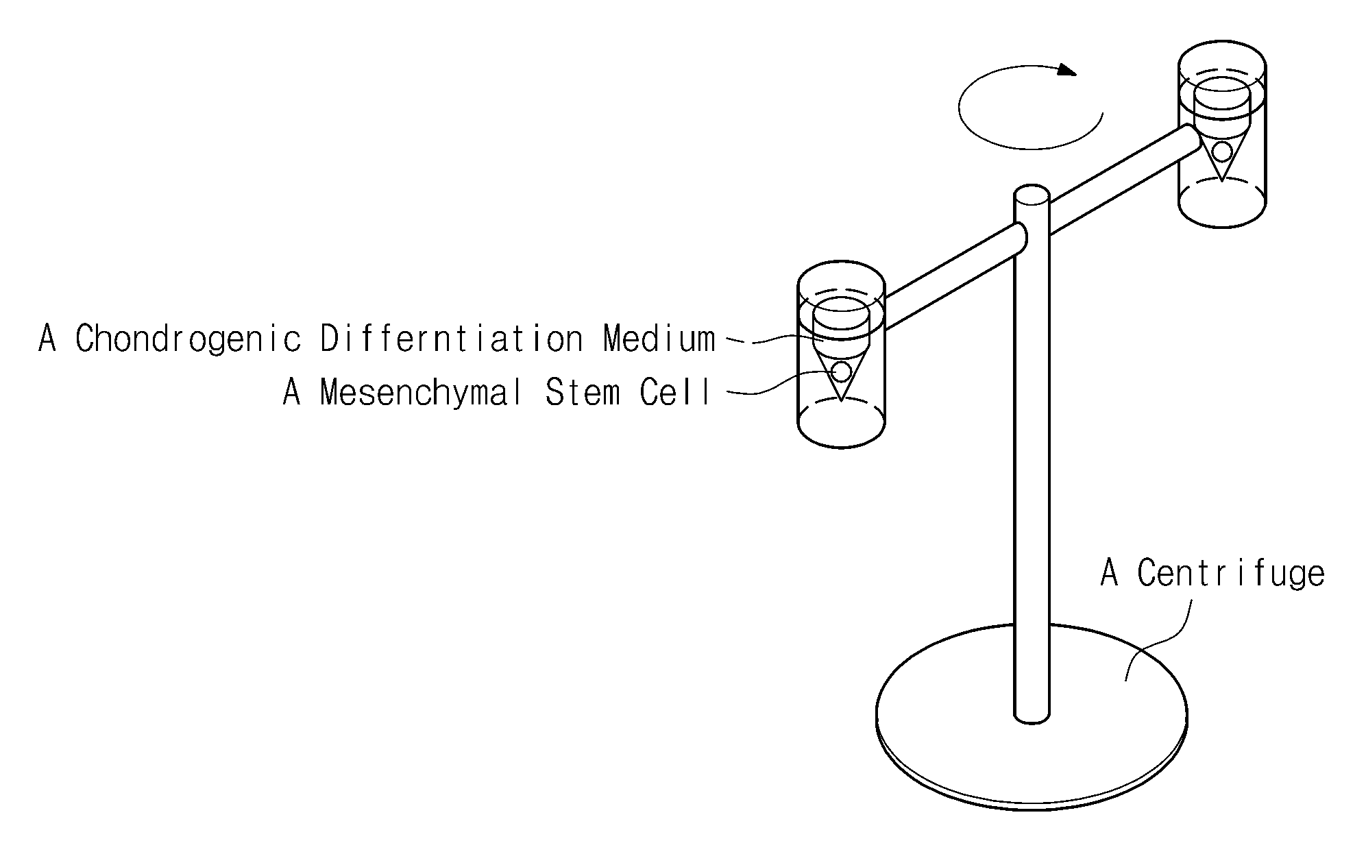
[0041]Each of the three-dimensional structures thus manufactured was placed in a chondrogenic differentiation medium [high glucose DMEM (hyclone, USA), ITS (ITS LIQUID MEDIA SUPPLEMENT) (Sigma, USA), 50 μg / ml ascorbic acid 2-pho...

Example

Experimental Example 4

Immunohistological Analysis 1

[0044]Each of the mesenchymal stem cells incubated in Experimental Examples 3-1 and 3-2 and included in the PGA scaffolds was washed respectively with PBS (Phosphate Buffer Saline) and fixed in a 10% formalin solution. Each of the fixed samples was dehydrated and embedded in paraffin blocks. After the blocks were cut into 4 μm sections, some of the sections were stained with safranin-O, and microscopic photos (depicted respectively in FIGS. 2a and 2b) were captured. GAG (glycosaminoglycan) protein, a marker for expression of aggrecan genes which are characteristic of chondrogenic cells, is stained red with safranin-O. Comparing the photo of the mesenchymal stem cells from the group subjected to centrifugal force in FIG. 2b with that of the mesenchymal stem cells of the control group (in FIG. 2a) that were incubated in a static state without being subjected to centrifugal force, it is apparent from the increased number of red-staine...


PUM


Abstract

Provided are a method of chondrogenic differentiation from mesenchymal stem cells and a composition comprising chondrogenic cells differentiated using the method to treat diseases caused by cartilage damage. In the method, a centrifugal force is applied to human mesenchymal stem cells to be differentiated into chondrogenic cells. The chondrogenic differentiation may be achieved at moderate cost without using expensive cytokines or growth factors by periodically applying only a centrifugal force. According to the method of chondrogenic differentiation from mesenchymal stem cells in the present embodiment, chondrogenic cells may be also differentiated from human mesenchymal stem cells.

Description

CROSS-REFERENCE TO RELATED APPLICATIONS[0001]This U.S. non-provisional patent application claims priority under 35 U.S.C. § 119 of Korean Patent Application No. 10-2008-0120190, filed on Nov. 29, 2008, the entire contents of which are hereby incorporated by reference.BACKGROUND OF THE INVENTION[0002]The present invention disclosed herein relates to a method of chondrogenic differentiation from mesenchymal stem cells, and a composition comprising chondrogenic cells differentiated using the method to treat disease caused by cartilage damage.[0003]Stem cells are cells before being differentiated into individual cells constituting a tissue. Stem cells refer to cells that can proliferate infinitely in an undifferentiated state and have the potential to differentiate into various types of tissue cells through specific differentiation stimuli.[0004]According to their differentiation potential, stem cells can be divided into embryonic stem cells (ESCs) and adult stem cells (ASCs) (also know...

Claims


Application Information

Patent Timeline
IPC IPC(8): A61K35/12C12N5/06C12N5/08
CPCA61K35/12C12N5/00C12N2525/00C12N2506/21C12N5/0655C12N5/0602
Inventor JEONG, MIN-SUKJUNG, MOON-YOUNPARK, SEON-HEE
Owner ELECTRONICS & TELECOMM RES INST
Why Eureka
Try Eureka