System and catheter for image guidance and methods thereof

a technology of ultrasound imaging and system, applied in the field of ultrasound imaging catheters, can solve the problems of atrio-esophageal fistula and thrombosis, the formation of atrio-esophageal fistulas and thrombosis, and the limitations of existing technologies

Active Publication Date: 2010-06-17
ACIST MEDICAL SYST
View PDF38 Cites 121 Cited by
  • Summary
  • Abstract
  • Description
  • Claims
  • Application Information

AI Technical Summary

Benefits of technology

[0015]The linear translation control system may be arranged to impart continuous controlled translation to the ultrasonic imaging core. The linear translation control system may be arranged to impart controlled bidirectional translation to the ultrasonic imaging core.

Problems solved by technology

AF ablation procedures are however not without serious risks.
Such risks include the formation of atrio-esophageal fistulas and thrombus.
These existing technologies however have important limitations.
Fluoroscopy provides a two-dimensional view of external structures, has poor soft tissue contrast, requires the injection of contrast, and exposes the patient and staff to radiation.
However, EAM is time consuming, complex, and does not provide real-time imaging of cardiac tissue.
Further, registration can be lost due to cardiac motion.
However, ICE catheters are practically limited to imaging from the right atrium due to their relatively large size.
This leads to relatively poor image performance, particularly for key cardiac structures such as the left pulmonary veins.
However, relatively poor near-field resolution of current TEE probes makes imaging of important cardiac structures difficult, such as the left atrium posterior wall.
Further, TEE often requires general anesthesia due to patient discomfort from large probe size.

Method used


Image

  • System and catheter for image guidance and methods thereof
  • System and catheter for image guidance and methods thereof
  • System and catheter for image guidance and methods thereof

Examples


Embodiment Construction

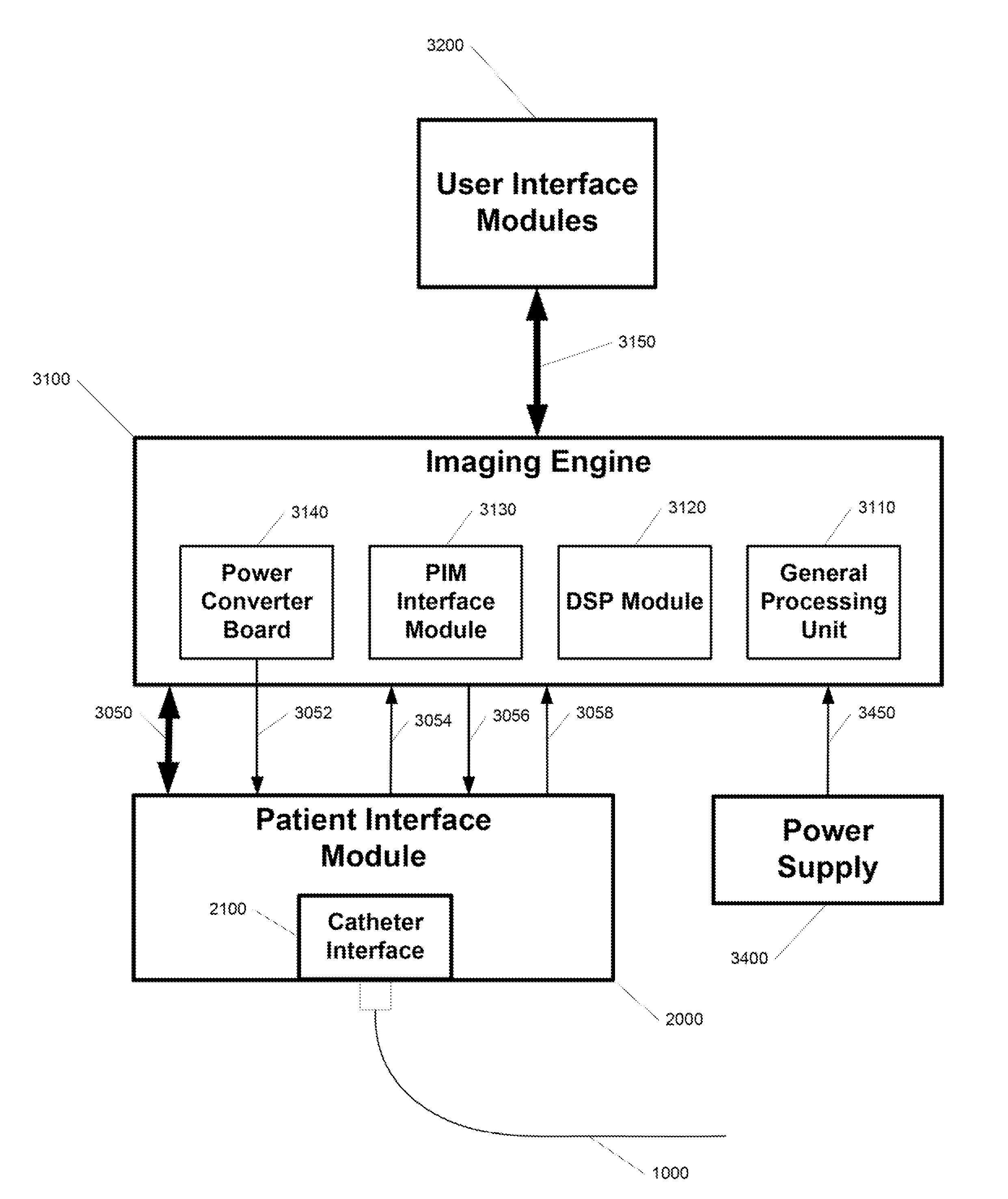
[0045]FIG. 1 shows a high-level diagram of an echocardiographic system and catheter. The system comprises an imaging engine 3100 and a patient interface module (PIM) 2000. The imaging engine 3100 is the central component of the system and performs all image generation, display, and control of the system components. The imaging engine 3100 comprises a general processing unit 3110, a digital signal processing (DSP) module 3120, and a PIM interface module 3130. The PIM 2000 is in mechanical and electrical communication with an echocardiographic catheter 1000.

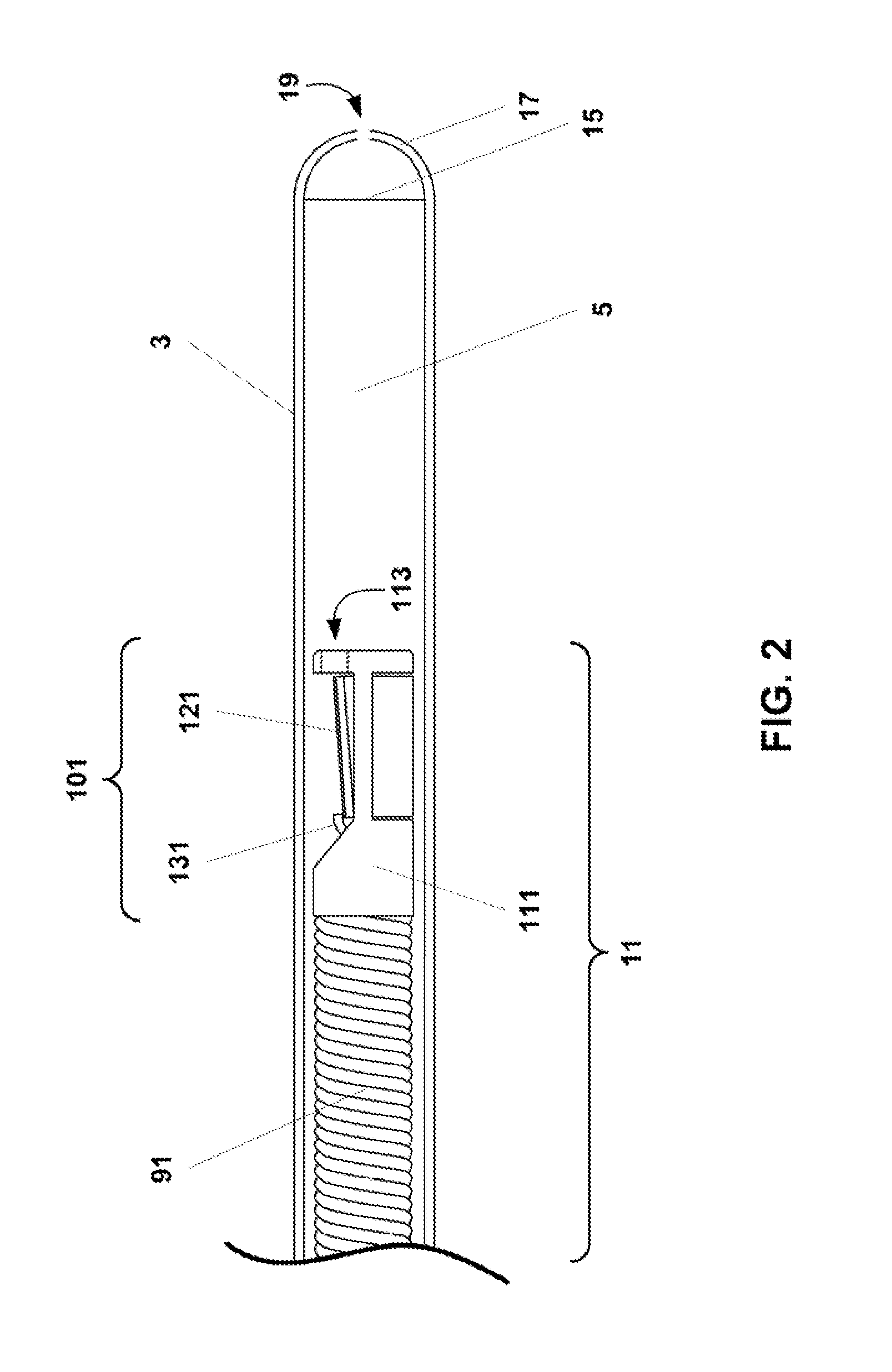
[0046]A catheter is a common medical device comprising a flexible tubular body having a proximal end and a distal end. A catheter configured in accordance with an embodiment of the present invention may comprise an outer tube having a proximal end, an inner sheath slidingly received within the outer tube and extending distally from the outer tube, and a rotatable shaft (or drive cable) extending from the proximal end of the outer t...


PUM


Abstract

A catheter-based imaging system comprises a catheter having a telescoping proximal end, a distal end having a distal sheath and a distal lumen, a working lumen, and an ultrasonic imaging core. The ultrasonic imaging core is arranged for rotation and linear translation. The system further includes a patient interface module including a catheter interface, a rotational motion control system that imparts controlled rotation to the ultrasonic imaging core, a linear translation control system that imparts controlled linear translation to the ultrasonic imaging core, and an ultrasonic energy generator and receiver coupled to the ultrasonic imaging core. The system further comprises an image generator coupled to the ultrasonic energy receiver that generates an image.

Description

PRIORITY CLAIM[0001]The present application claims the benefit of copending U.S. Provisional Patent Application Ser. No. 61 / 120,762, filed Dec. 8, 2008, and U.S. Provisional Patent Application Ser. No. 61 / 158,075, filed Mar. 6, 2009, which applications are incorporated herein by reference in their entirety.BACKGROUND[0002]The present invention generally relates to ultrasound imaging catheters. The present invention further relates to transesophageal echocardiographic catheters for the purpose of guiding cardiac interventions. The present invention still further relates further to intracardiac echocardiographic catheters for the purpose of guiding cardiac interventions.[0003]Atrial fibrillation (AF) is the most common arrhythmia in the United States and other developed countries. Nearly two (2) million individuals in the United States have AF. Catheter ablation is increasingly selected therapy for AF. Radiofrequency ablation is the most common form of AF ablation.[0004]AF ablation pr...

Claims


Application Information

Patent Timeline
Patent Type & Authority Applications(United States)
IPC IPC(8): A61B8/14
CPCA61B8/12A61B8/445A61B8/4461A61B1/018A61B8/0841A61B8/5223A61B8/5253A61B18/1492A61B2017/00243A61B2017/00557A61B34/25A61B90/37A61B2090/3784G16H50/30A61B1/00082A61B1/233A61B1/2733A61B1/3137A61B8/0883A61B8/4466A61B8/4494A61B2017/00991A61B2018/00577A61B2018/00982A61B2018/0212
Inventor MOORE, THOMAS C.WATERS, KENDALL R.BUECH, STEPHANIE J.ZELENKA, ROBERT
Owner ACIST MEDICAL SYST
Why Eureka
Try Eureka