Use of cyanine dyes for the diagnosis of proliferative diseases

a technology of proliferative diseases and cyanine dyes, which is applied in the field of using cyanine dyes for the diagnosis of proliferative diseases, can solve the problems of mammography being associated with a significant and cumulative risk of radiation exposure, cancer financial burden, and affecting the quality of life of patients,

Inactive Publication Date: 2010-07-01
SCHERING AG
View PDF11 Cites 32 Cited by
  • Summary
  • Abstract
  • Description
  • Claims
  • Application Information

AI Technical Summary

Benefits of technology

[0015]The ability of the hydrophilic compounds usable according to the present invention and in particular of the cyanine dyes SF64, 5-29, 5-36, and 5-41 to act as contrast agent is in part determined by oxy- and dioxy-hemoglobin concentrations, blood oxidant saturation, contrast agent uptake into tissue and organical concentration, however, it is possible to increase the specificity and / or sensitivity of the compounds usable according to the present invention and in particular of SF64, 5-29, 5-36, and 5-41 by coupling it to a targeting compound which binds specifically to structures which are preferentially or exclusively present on proliferating cells and tissues or in the vicinity of proliferating cells and tissue. Some of these structures are associated directly with the proliferating cell or are associated with cells in the vicinity of the proliferative tissue. The former are structures altered or over-expressed in the proliferating cell like, for example, growth factor receptors, like somatostatin receptor or epidermal growth factor receptor (FGFR). A large variety of such structures have been identified by now and comprise without limitation growth factor receptors, G-protein coupled receptors, pore proteins, ion channels, drug efflux pumps, accessory binding sites for growth factors, heparan sulfate, membrane bound proteases, adhesion molecules, T cell receptors and selectins, in particular EGF, TGF, CEA, Lewis Y, CD 20, CD 33, or CD38. Other structures, which can be targeted are T-cell-defined cancer-associated antigens belonging to unique gene products of mutated or recombined cellular genes, in particular cyclin-dependent kinase 4 (CDK4), p15Ink4b, p53, AFP, β-catenin, caspase 8, p53, p21Ras mutations, Bcr-abl fusion product, MUM-1 MUM-2, MUM-3, ELF2M, HSP70-2M, HST-2, KIAA0205, RAGE, myosin / m, 707-AP, CDC27 / m, ETV6 / AML, TEL / Aml1, Dekcain, LDLR / FUT, Pm1-RARα, TEL / AMLI; Cancer-testis. (CT) antigens, in particular NY-ESO-1, members of the MAGE-family (MAGE-A1, MAGE-A2, MAGE-A3, MAGE-A4, MAGE-A6 MAGE-10, MAGE-12), BAGE, DAM-6, DAM-10, members of the GAGE-family (GAGE-1, GAGE-2, GAGE-3, GAGE-4, GAGE-5, GAGE-6, GAGE-7B, GAGE-8), NA-88A, CAG-3, RCC-associated antigen G250; Tumor virus antigens, in particular human papilloma virus (HPV)-derived E6 or E7 oncoproteins, Epstein Barr virus EBNA2-6, LMP-1, LMP-2; overexpressed or tissue-specific differentiation antigens, in particular gp77, gp100, MART-1 / Melan-A, p53, tyrosinase, tyrosinase-related protein (TRP-1 and TPR-2), PSA, PSM, MC1R; widely expressed antigens, in particular ART4, CAMEL, CEA, CypB, HER2 / neu, hTERT, hTRT, ICE, Muc1, Muc2, PRAME RU1, RU2, SART-1, SART-2, SART-3, and WT1.
[0036]The use of the present invention can be for routine diagnosis i.e. for screening for the respectively indicated diseases. However, in a further embodiment the conjugates are used once the disease has been diagnosed with, for example, a standard x-ray procedure, e.g. mammography, a whole body scan or MRI. The patient is then examined for metastases and / or small (additional) primary tumor(s). Such an examination can occur for a better assessment of the severity, e.g. stage of the disease, or to determine the best treatment options and / or prior, during and / or after a treatment procedure (e.g. drugs, radiation or surgery). If performed prior to a treatment procedure the use of present invention due to its high sensitivity allows the determination whether, e.g. metastases have already formed in the vicinity of the primary tumor, and, thus, a better determination of the treatment regimen, e.g. whether a lumpectomy or rather a mastectomy is indicated in breast cancer.
[0037]After treatment the use of the diagnostic procedure of the present invention allows to assess the success of the treatment procedure and to determine subsequent treatment regiments, e.g. radiation or chemotherapy. When used during a surgical procedure it is, for example, possible to detect metastases in tissue, e.g. lymph nodes, surrounding of the primary tumor. In this embodiment the use of the present invention allows more complete removal of tumors or metastases during a surgical procedure.

Problems solved by technology

The financial costs of cancer are overwhelming.
However, mammography has been associated with a significant and cumulative risk of radiation exposure in particular in premenopausal woman, which have denser breast tissue and require higher radiation dosages as older woman to obtain a sufficient sensitivity.
Mammography has also been criticized for the forceful manipulation of the breast during the procedure, which might facilitate dissemination of tumor cells.
MRI imaging due to its high spatial resolution has a vastly superior sensitivity in comparison to x-ray based imaging techniques like mammography, however, it is less specific (specificity ranging from 37% to 97% and the predictive value for woman not previously diagnosed with breast cancer is less than 2%) much more expensive and time consuming and, thus, less amenable to mass screening of patients.
Transillumination, however, had low spatial resolution and afforded little in spectral quantification of the lesions detected.
Hence, transillumination did not attain sufficient sensitivity and specificity to be used clinically.

Method used


Image

  • Use of cyanine dyes for the diagnosis of proliferative diseases
  • Use of cyanine dyes for the diagnosis of proliferative diseases
  • Use of cyanine dyes for the diagnosis of proliferative diseases

Examples


examples

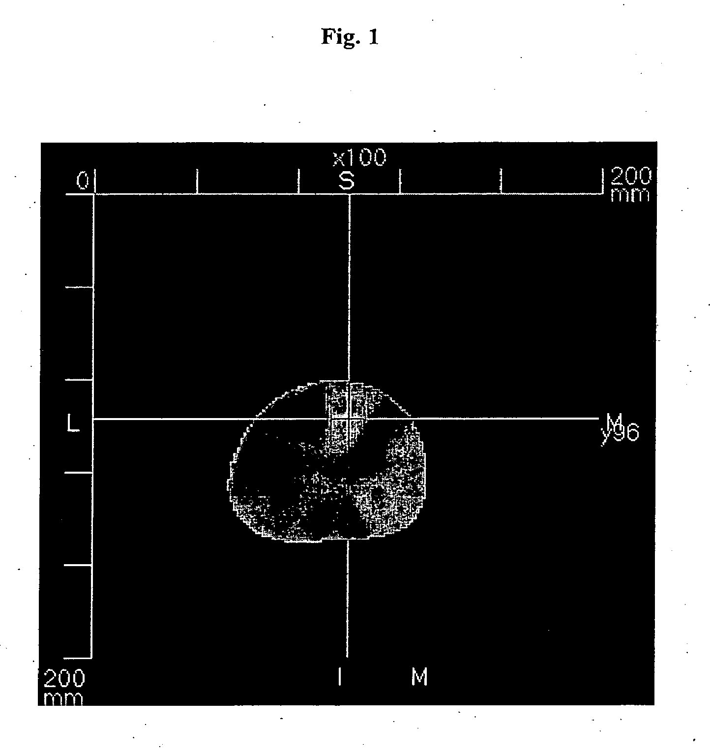
Optical Mammography after i.v. Injection of SF64 in a Female Patient with Invasive Breast Cancer in the Right Breast

[0046]A dose of 0.1 mg / kg body weight of SF64 was, after dilution of the lyophilisate with 0.9% normal saline, injected intravenously via an indwelling forearm canula. The patient was imaged with an optical computed tomographic laser mammography device in a prone position with her breast hanging freely into the imaging chamber, being surrounded by air. Image acquisition was started 1 hour 10 minutes after i.v. injection of SF64, and image reconstruction was performed using the fluorescence mode, thus almost exclusively showing the fluorescence signal of SF64. The contrast dye did accumulate in this invasive breast cancer, which is represented in the image by the bright spot. This led to the marked fluorescence signal of SF64 in the image.

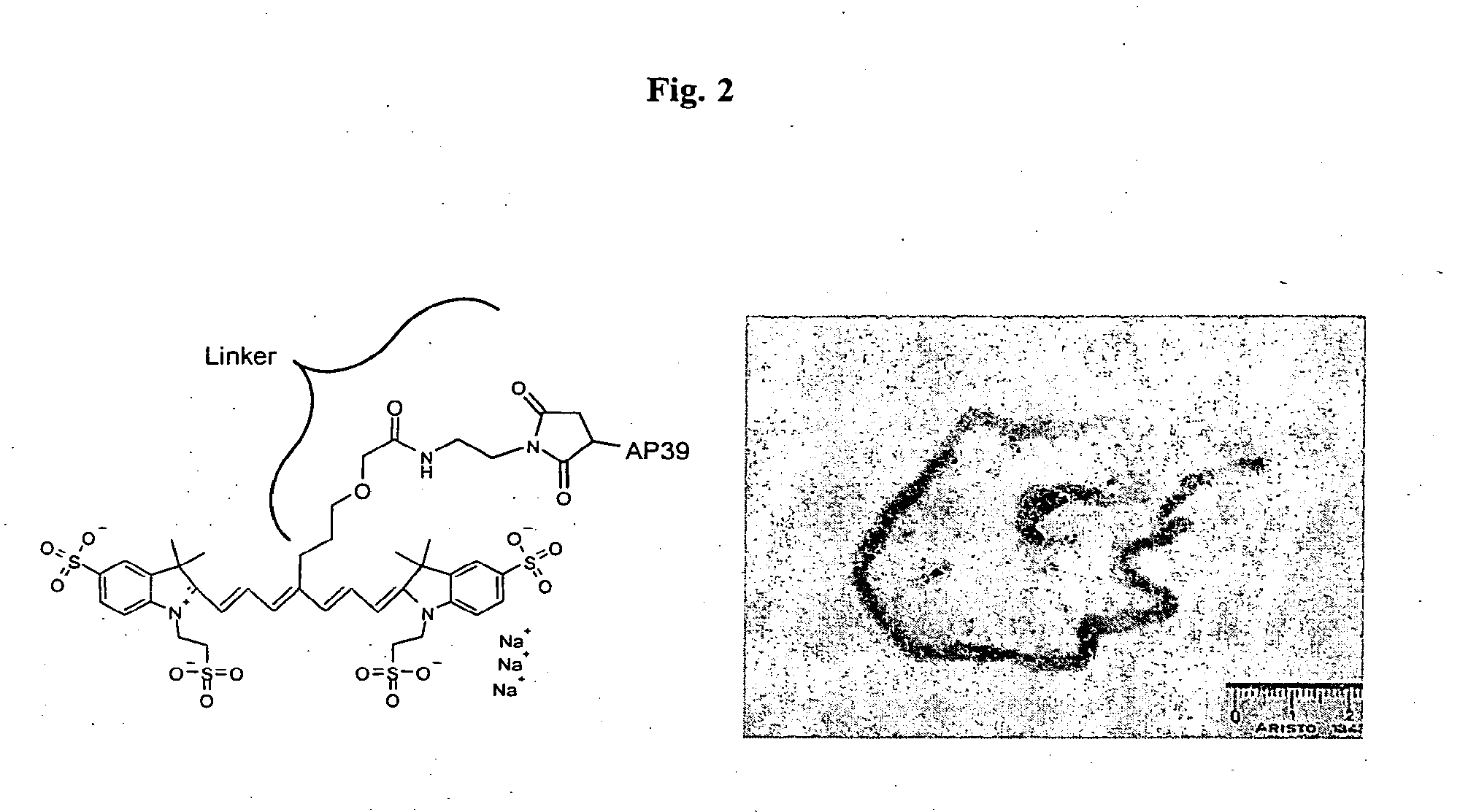
Determination of Fluorescence Quantum Yield of Several Tricarbocyanine Dyes

[0047]Compounds 6-4, 5-36, 5-29, 5-41, NIR96009, NIR96005 ...


PUM

PropertyMeasurementUnit
sizeaaaaaaaaaa
diameteraaaaaaaaaa
sizeaaaaaaaaaa

Abstract

The present invention concerns the use of the cyanine dye SF64 for the diagnosis of proliferative diseases upon administration of less than 5 mg / kg body weight.

Description

[0001]The present invention concerns the use of cyanine dyes, in particular SF64, 5-29, 5-36, and / or 5-41 for the diagnosis of proliferative diseases, in particular tumor diseases upon administration of less than 0.1 mg / kg body weight.BACKGROUND OF THE INVENTION[0002]Cancer is the second leading cause of death among Americans and is responsible for one out of every four deaths in the United States. In 2004 over 560,000 Americans or more than 1,500 people a day will die of cancer. Over 18 million new cases of cancer have been diagnosed since 1990 and about 1.4 million new cases will be diagnosed in 2004 alone. This estimate does not include preinvasive cancer or more than 1 million cases of none-melanoma-skin cancer expected to be diagnosed this year. The financial costs of cancer are overwhelming. According to the National Institute of Health cancers cost in the United States are more than $ 189 billion in 2003. This amount includes over $ 64 billion in direct medical costs and more...

Claims


Application Information

Patent Timeline
Patent Type & Authority Applications(United States)
IPC IPC(8): A61K49/00
CPCA61K49/0032A61K49/0058
Inventor LICHA, KAISCHIRNER, MICHAELPESSEL, MARTINBAHNER, MALTE
Owner SCHERING AG
Why Eureka
Try Eureka