Needle and tine deployment mechanism

a deployment mechanism and needle technology, applied in the field of medical devices and methods, can solve the problems of increasing the stress imparted to the needle, the ability to increase the exit angle, for example, by lengthening the ramp, and the ramp deflecting, etc., so as to improve the geometry and stabilize the effect of the geometry, the effect of gradual bending radius

Active Publication Date: 2010-09-02
GYNESONICS
View PDF31 Cites 71 Cited by
  • Summary
  • Abstract
  • Description
  • Claims
  • Application Information

AI Technical Summary

Benefits of technology

[0072]By proximally retracting and radially offsetting the axis of the spike 210 relative to the central electrode port 214 and the central axis of the central member 202, an improved and stabilized geometry is provided for deploying central electrode 230, as illustrated in FIGS. 20A-20C. Initially, as shown in FIG. 20A, the central electrode 230 is retracted within the axially aligned electrode advancement passage 205 with a distal, tissue-penetrating tip 232 of the electrode positioned proximally of the central electrode deployment port 214. By advancing the central electrode 230, using the mechanisms described with previous embodiments, distal tip 232 will be distally advanced into tissue along a generally linear path, as shown in FIG. 20B. The stability and alignment of the needle electrode 230 is partially maintained by traveling through the channel or groove 216 on the inner surface of the introducer tip 210.
[0073]After the device 200 has been introduced into tissue and the central electrode 230 and optionally peripheral electrodes (not shown) are advanced into the tissue, the central member 202 can sometimes become misaligned so that the electrode 230 is stressed and bent at the region where it exits from the axially aligned electrode advancement passage 205, as shown in FIG. 20C. By having a rounded peripheral edge 218 surrounding the central electrode port 214, the stress on the needle can be relieved by allowing a more gradual bending radius. Moreover, the rounded peripheral edge also facilitates retracting the electrode 230 as binding and friction are reduced.
[0074]Referring now to FIGS. 21A-21C, a further embodiment of the central member 300 terminates in a spike 302 having a trocar cutting surface 304 and an opposed chamfered surface 306. An electrode transition cavity 308 is formed in the chamfered surface 306 and extends distally from a central electrode port 310, as best seen in FIGS. 21B and 21C. As illustrated, the cavity 308 has a generally ovoid shape, but it could also be spherical, conical, or have some other concave geometry. The exit port 310 may have a tapered geometry, as shown in FIG. 21B, or a more slotted geometry, as shown in FIG. 21C. In both cases, the transition of the central electrode from the central electrode passage through the electrode port 310 into the cavity 308 provides stress relief both as the electrode is advanced and as the central member 300 is manipulated during use. Preferably, the deployment device of FIGS. 21A-21C will include the peripheral electrode channels 312 similar to those described in previous embodiments.

Problems solved by technology

While such angles of deflection could theoretically be obtained by providing relatively short, sharply angled ramps, such short, steeply deflected ramps will increase the stress imparted to the needles.
The ability to increase the exit angle, for example, by lengthening the ramp, however, can be limited in certain designs, particularly designs having a relatively small diameter central member.

Method used


Image

  • Needle and tine deployment mechanism
  • Needle and tine deployment mechanism
  • Needle and tine deployment mechanism

Examples


Embodiment Construction

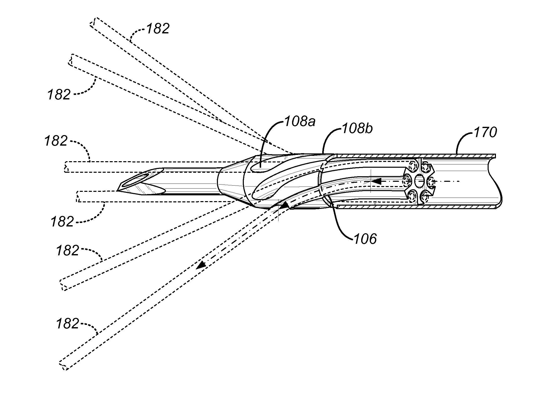
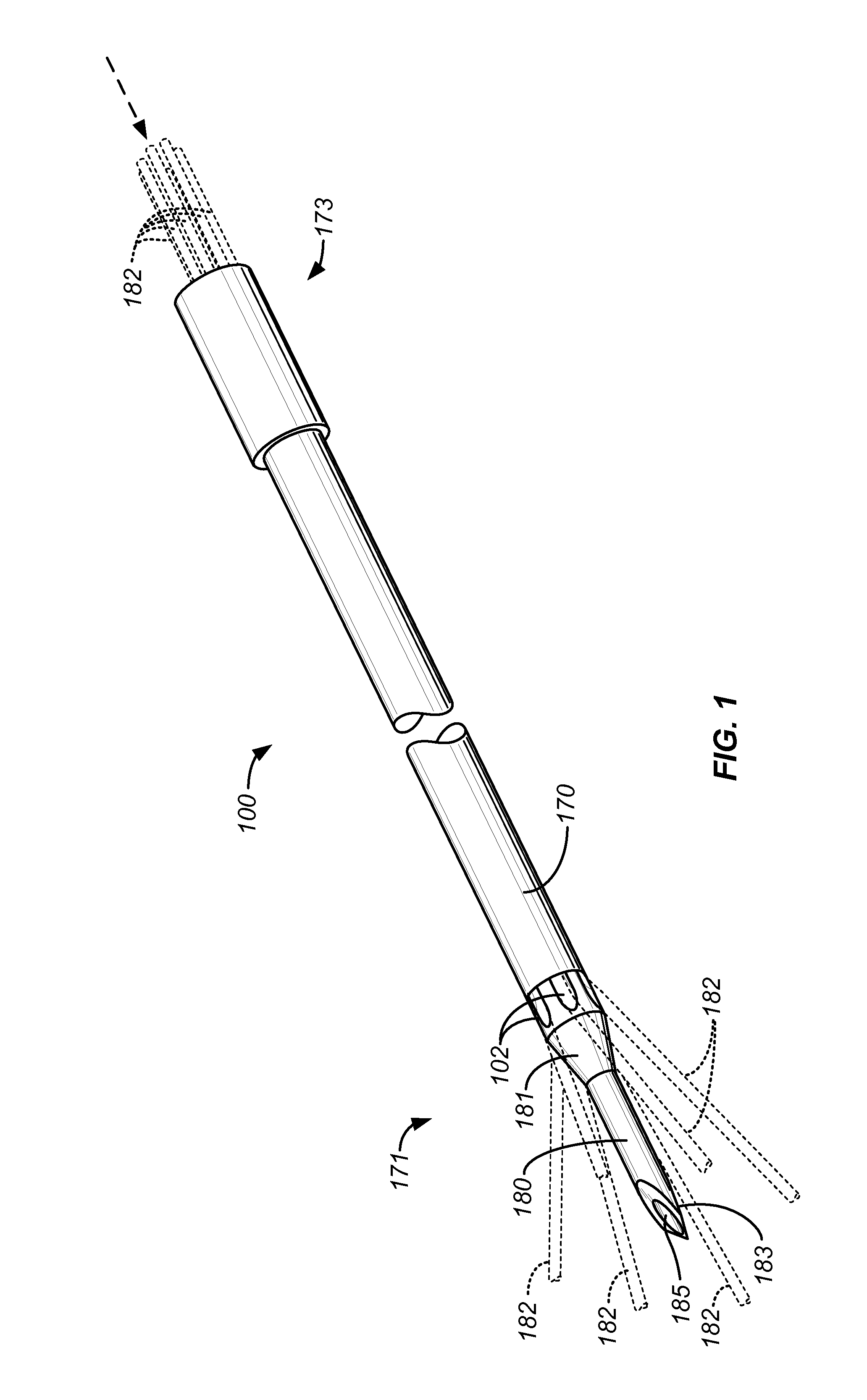
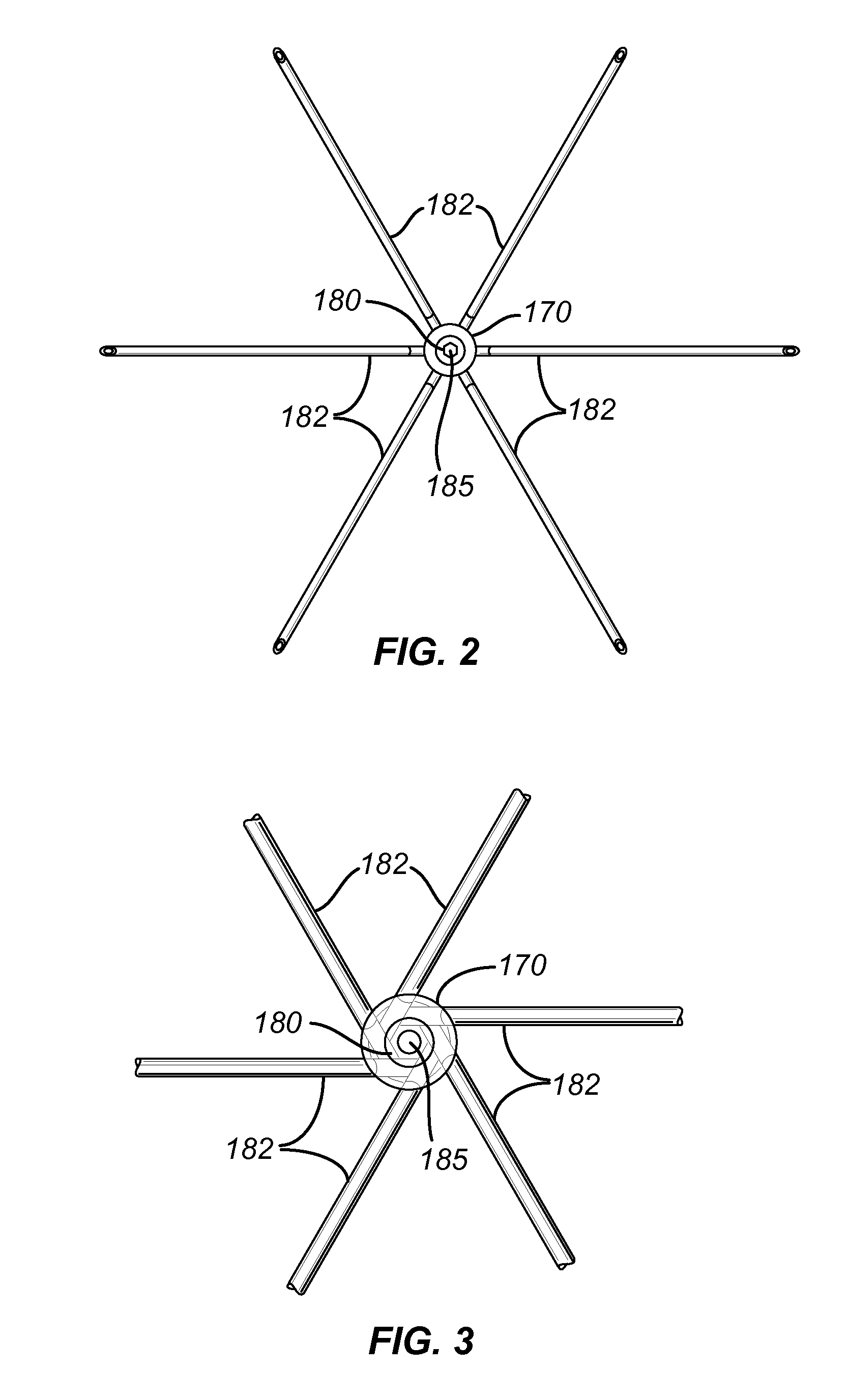
[0050]The present invention provides electrode deployment structures comprising needle electrode deployment shafts capable of reciprocatably deploying a plurality of needle electrodes into solid tissue. The needle electrode deployment shafts will comprise a central member having a proximal end, a distal end, and a longitudinal axis therebetween. Typically, the central member will have a distal end which is configured to permit self-penetration, e.g. the central member may itself be a needle having a sharpened or chamfered tip which permits the central member to be advanced into tissue by simply pushing. In other embodiments, the central member could comprise a hollow tubular body, commonly referred to as a cannula, having a needle or stylet removably received within a central lumen thereof. The cannula could then be introduced by placing the stylet with its sharpened tip extending from the distal end of the cannula and pushing the assembly of the cannula and stylet into tissue. The ...


PUM


Abstract

A needle electrode deployment shaft includes a central member and a plurality of needle electrodes. The central member has a plurality of needle advancement channels formed therein. The needle electrodes are disposed within the advancement channels and each advancement channel terminates in a ramp portion which deflects the needles radially outwardly as they are axially advanced. The ramps may be spirally or acutely configured in order to increase the distance through which the needles may be bent as they are axially advanced. Additionally, the central member may have a radially reduced distal tip in order to decrease tissue insertion forces.

Description

CROSS-REFERENCES TO RELATED APPLICATIONS[0001]The present application claims the benefit of provisional application 61 / 156,270 (Attorney Docket No. 025676-002300US), filed on Feb. 27, 2009, and is related to but does not claim the benefit of provisional patent application No. 61 / 091,708 (Attorney Docket No. 025676-002200US), filed on Aug. 25, 2008, the full disclosures of which are incorporated herein by reference.BACKGROUND OF THE INVENTION[0002]1. Field of the Invention[0003]The present invention relates generally to medical devices and methods. More particularly, the present invention relates to a therapy device having a deployable treatment needle with a plurality of tines deployable in a straight path from said needle.[0004]Uterine fibroids are benign tumors in the uterine wall and are the most common tumor of the female pelvis. Fibroids afflict up to 30% of women of childbearing age and can cause significant symptoms including discomfort, pelvic pain, mennorhagia (excessive bl...

Claims


Application Information

Patent Timeline
Patent Type & Authority Applications(United States)
IPC IPC(8): A61B19/00A61B8/00A61B18/18
CPCA61B18/1477A61B2018/00547A61B2018/00559A61B2018/00577A61B2019/528A61B2018/143A61B2018/1475A61B2019/5263A61B2018/1427A61B2034/2063A61B2090/3784
Inventor PLACEK, BRIANDECKMAN, ROBERT K.
Owner GYNESONICS
Why Eureka
Try Eureka