Information processing apparatus for registrating medical images, information processing method and program

a technology of information processing apparatus and medical images, applied in the field of information processing apparatus for registering medical images, information processing method and program, can solve the problems of high signal to noise ratio, heavy work load at the time of imaging, and inability to obtain detailed information of the interior portion of a human body, etc., and achieve high speed and high precision.

Active Publication Date: 2010-09-23
CANON KK
View PDF7 Cites 80 Cited by
  • Summary
  • Abstract
  • Description
  • Claims
  • Application Information

AI Technical Summary

Benefits of technology

[0029]According to the present invention, an ultrasonic image and three-dimensional medical image can be registered with high precision and at high speed.

Problems solved by technology

On the other hand, since information of the radiation direction of X-rays is lost, detailed information of the interior portion of a human body cannot be obtained.
On the other hand, at the time of imaging by the MRI, a contrast agent has to be normally applied to a human body, resulting in a heavy work load at the time of imaging.
On the other hand, an image (ultrasonic image) of the ultrasonic imaging apparatus has a high signal to noise ratio, which may often disturb diagnosis.
However, in order to register the ultrasonic image and three-dimensional medical image, some problems have to be solved.

Method used


Image

  • Information processing apparatus for registrating medical images, information processing method and program
  • Information processing apparatus for registrating medical images, information processing method and program
  • Information processing apparatus for registrating medical images, information processing method and program

Examples


first embodiment

1. Functional Arrangement of Information Processing Apparatus

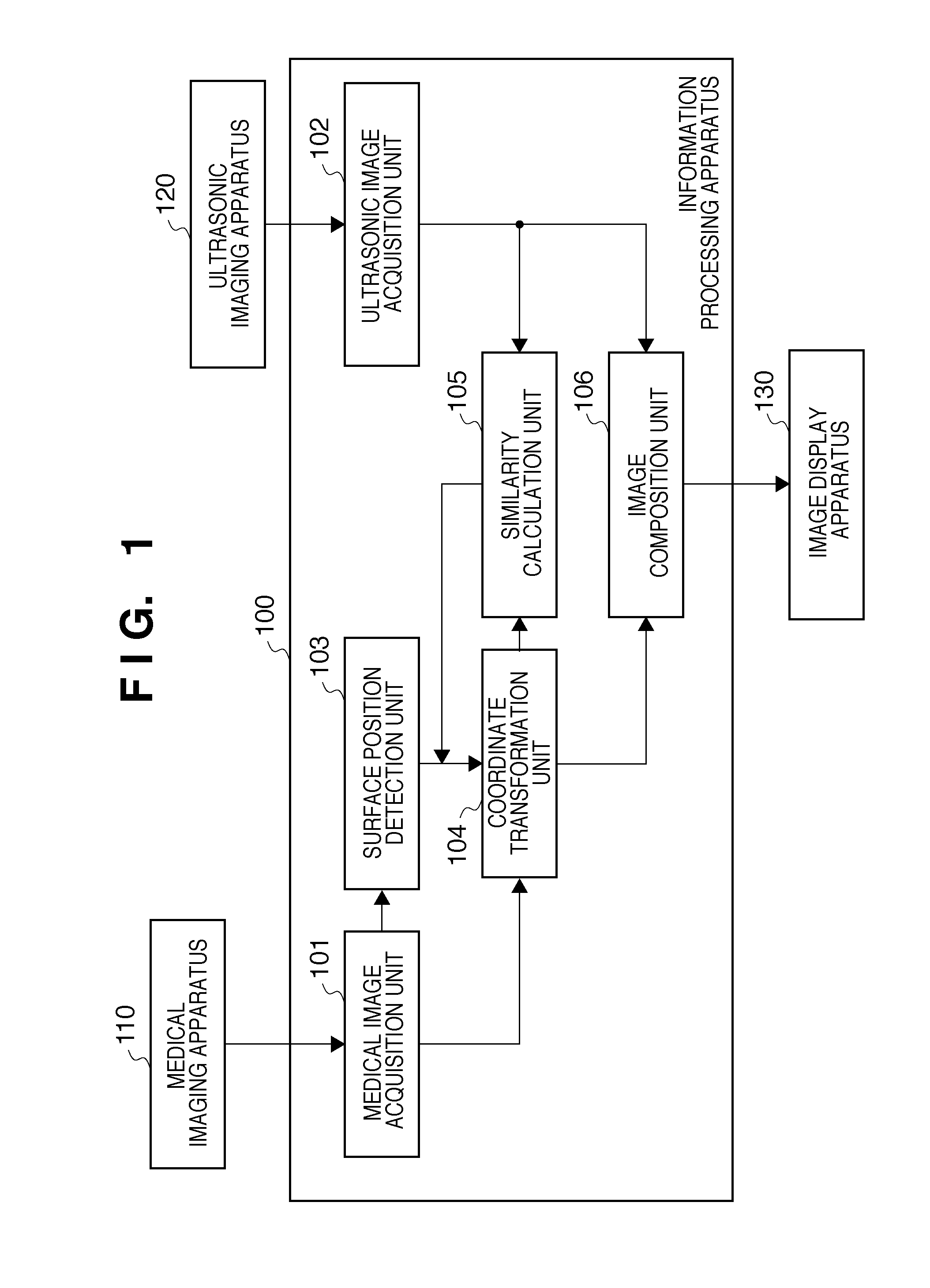
[0057]FIG. 1 is a block diagram showing the functional arrangement of an information processing apparatus 100 according to this embodiment. The information processing apparatus 100 is connected to a medical imaging apparatus 110 and ultrasonic imaging apparatus 120, and executes processing for registering images of a target region captured by the connected apparatuses. Note that the registration is to transform the coordinates of any of images so that pieces of image information in the images of the target region captured by the respective apparatuses match.

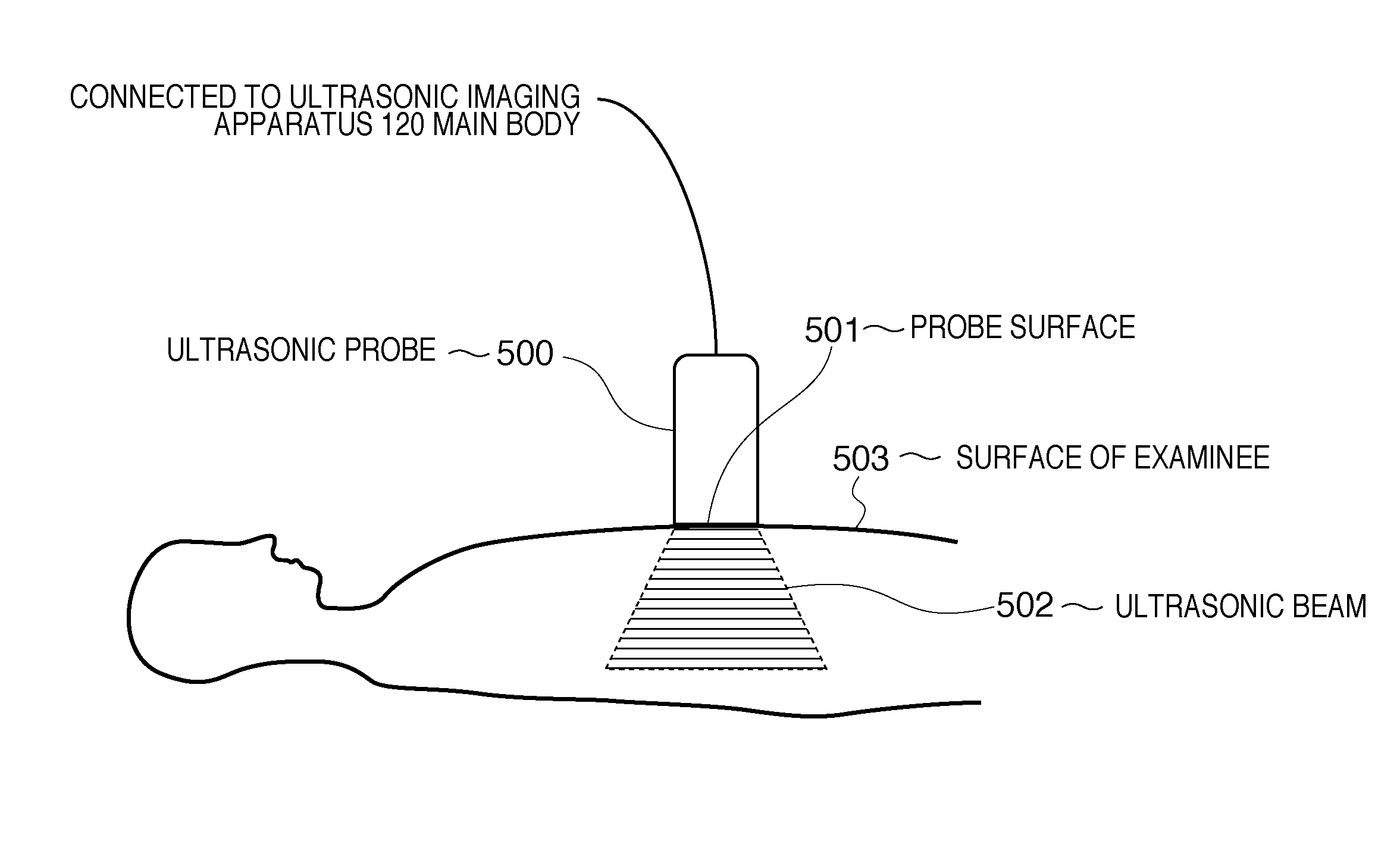
[0058]The medical imaging apparatus 110 is an apparatus such as an X-ray CT, MRI, SPECT, or PET, which captures a medical image of the interior portion of an examinee (object). The ultrasonic imaging apparatus 120 is an apparatus which captures an image of a second region of the interior portion of the examinee via ultrasonic waves by bringing an ultrasonic probe (imagi...

second embodiment

[0201]In the first embodiment, an ultrasonic image and medical image are registered based on pieces of image information in these images. However, the present invention is not limited to this. Coordinate transformation parameters may be calculated using measurement results obtained by measuring the position and orientation of an ultrasonic probe in addition to the image information. This embodiment will be described in detail below.

[0202]

[0203]FIG. 11 is a block diagram showing the functional arrangement of an information processing apparatus 1100 according to this embodiment. The information processing apparatus 1100 is connected to a medical imaging apparatus 1110, ultrasonic imaging apparatus 1120, and position and orientation measurement device 1140.

[0204]The medical imaging apparatus 1110 is an apparatus such as an X-ray CT or MRI, which captures a medical image of the interior portion of an examinee. The ultrasonic imaging apparatus 1120 is an apparatus which captures an image...


PUM


Abstract

An information processing apparatus to register an ultrasonic image and a three-dimensional medical image at high speed is disclosed. The information processing apparatus includes: a medical image acquisition unit which acquires a medical image captured by a medical imaging apparatus in which an imaging unit captures an image of an object at a non-contact position with respect to the object, an ultrasonic image acquisition unit which acquires an ultrasonic image captured by an ultrasonic imaging apparatus in which an ultrasonic probe captures an image of the object at a position in contact with a surface of the object, and a coordinate transformation unit which transforms coordinates of the medical image or ultrasonic image with reference to the contact position on the ultrasonic image, so that image information of the medical image matches that of the ultrasonic image.

Description

CROSS REFERENCE TO RELATED APPLICATIONS[0001]This application is a CONTINUATION of PCT application No. PCT / JP2009 / 004637 filed on Sep. 16, 2009 which claims priority from the benefit of Japanese Patent Application No. 2008-311560 filed on Dec. 5, 2008, the disclosures of which are hereby incorporated by reference herein in their entirety.TECHNICAL FIELD[0002]The present invention relates to an information processing technique required to register images obtained by capturing images of an object using a plurality of imaging units.BACKGROUND ART[0003]In recent years, a doctor displays a captured medical image on a monitor and interprets the medical image on the monitor to make a diagnosis (image diagnosis) of a state or temporal change of a morbid portion of a patient.[0004]As an apparatus (medical imaging apparatus) for generating a medical image of this type, a plain radiographic apparatus, X-ray computed tomography apparatus (X-ray CT), nuclear magnetic resonance imaging apparatus ...

Claims


Application Information

Patent Timeline
Patent Type & Authority Applications(United States)
IPC IPC(8): G06K9/32
CPCA61B5/0095Y10S128/922A61B6/5229A61B6/5241A61B8/08A61B8/13A61B8/4245A61B8/5238G06T7/0024G06T7/2053G06T2207/10072G06T2207/10132G06T2207/30004A61B6/5247A61B6/03A61B8/4254G06T7/30G06T7/254
Inventor ISHIKAWA, RYOSATOH, KIYOHIDEUCHIYAMA, SHINJI
Owner CANON KK
Why Eureka
Try Eureka