Method for detecting abnormal spots of nucleic acid microarray

a nucleic acid microarray and abnormal spot technology, applied in the field of abnormal spot detection, can solve the problems of inability to detect chromosomal aberration, spot variation, and detection resolution, so as to reduce the variation of data among nucleic acid microarrays, accurate data, and reduced error in target nucleic acid labeled quantity valu

Inactive Publication Date: 2010-12-02
FUJIFILM CORP +1
View PDF0 Cites 3 Cited by
  • Summary
  • Abstract
  • Description
  • Claims
  • Application Information

AI Technical Summary

Benefits of technology

[0016]In the above method, by comparing labeled amount values of the abnormality detecting nucleic acids which are hybridized with different spots and considering a comparison value thereof, it becomes possible to detect abnormal spots and reduce variation of data among nucleic acid microarrays. Thereby, an error in the labeled quantity value of the target nucleic acid can be reduced, and more accurate data can be obtained.
[0017]Further, with the above quality inspection method, for example, a manufacturer of the nucleic acid microarray carries out hybridization with the nucleic acid microarray using a labeled abnormality detecting nucleic acid and provides the user with the resulting labeled quantity values. Then, the user can know which spots are the spots having problems in view of quality inspection by comparing the data with the labeled quantity value of the labeled abnormality detecting nucleic acid hybridized together with the labeled target nucleic acid of the nucleic acid microarray used in the test. Thereby, the user can easily carry out quality inspection for each spot on the nucleic acid microarray. In addition, since the quality inspection method uses a labeled quantity value of the labeled abnormality detecting nucleic acid used in the test for the quality inspection, it can be carried out with adding only slight steps (most of them can be dealt with a computer program), without requiring a special reagent for the quality inspection and without adding a special step for the quality inspection.

Problems solved by technology

However, this CGH method has a problem in detection resolution because it cannot detect chromosomal aberration if the chromosomal aberration does not occur within a large region of generally from 5 to 10 Mb or more (J. Inasawa and M. Mizukuchi, Rinsho Kensa (Clinical Inspection), 49, 497-502 (2005)).
There are cases in which the nucleic acid microarray produced by such a method is apt to cause variation in the amount of spot due to spotting error and the like and to generate an error of fluorescence signal caused by the gene to be measured.
In this method, there is a problem in that it requires staining, scanning, washing and the like complex operations.
Also, in order to use the nucleic acid microarray for the original purpose after staining the nucleic acid microarray, it is necessary to employ a washing step for removing the reacted dyestuff from the nucleic acid microarray and even the washing step is employed, it also poses a problem in that the dyestuff remains on the array.
Since the inspection is carried out not on all of the nucleic acid microarrays in this method, there is a possibility of finding that they are rejected articles at the stage of using them by the user.

Method used


Image

  • Method for detecting abnormal spots of nucleic acid microarray
  • Method for detecting abnormal spots of nucleic acid microarray
  • Method for detecting abnormal spots of nucleic acid microarray

Examples


example 1

Results of Analysis of Nucleic Acid Microarray in the Case of Eliminating Abnormal Spots

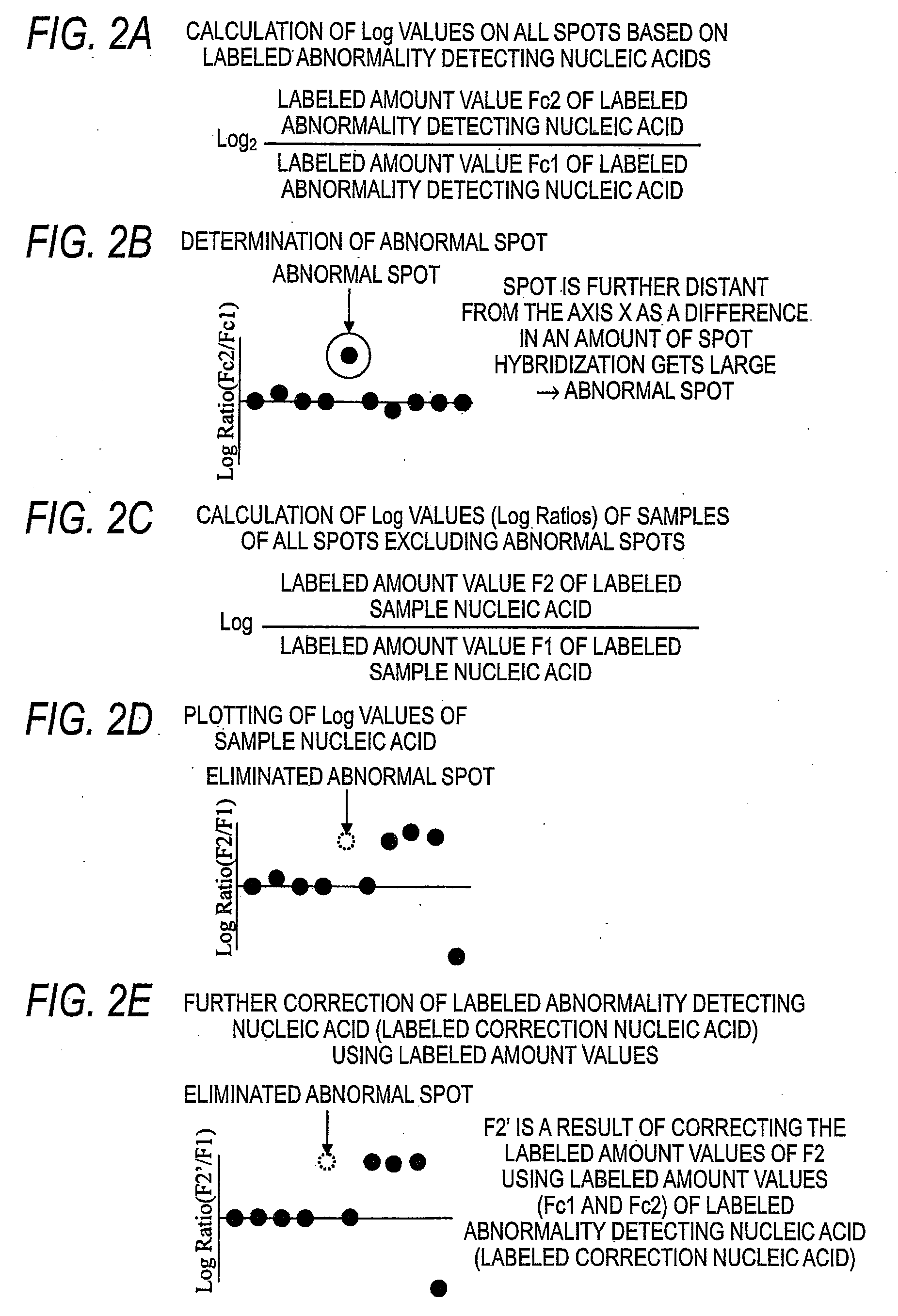
[0231]On the results of Comparative Example 1, abnormal spots were detected through numerical calculation by the following method.

[0232]By regarding the Cy5-Cot-1 DNA-derived fluorescence value obtained from the Cy3-Female-hybridized nucleic acid microarray as Fc1, and the Cy5-Cot-1 DNA-derived fluorescence value obtained from the Cy3-Male-hybridized nucleic acid microarray as Fc2, their Log Ratio {Log Ratio(Cot-1 / Cot-1)} was calculated as comparative value (DB) by the following formula.

Log Ratio(Cot-1 / Cot-1)=Log2(Fc2 / Fc1)

[0233]In this connection, the comparative value (DB) was calculated between spots having the same Block Number, the same Column Number and the same Row Number of the Cy3-Female-hybridized nucleic acid microarray and the y3-Male-hybridized nucleic acid microarray, and the comparative value (DB) was calculated on all of the spots spotted on the nucleic acid microarrays.

[0234]Next,...

example 2

Quality Inspection of Nucleic Acid Microarray Using Labeled Abnormality Detecting Nucleic Acid

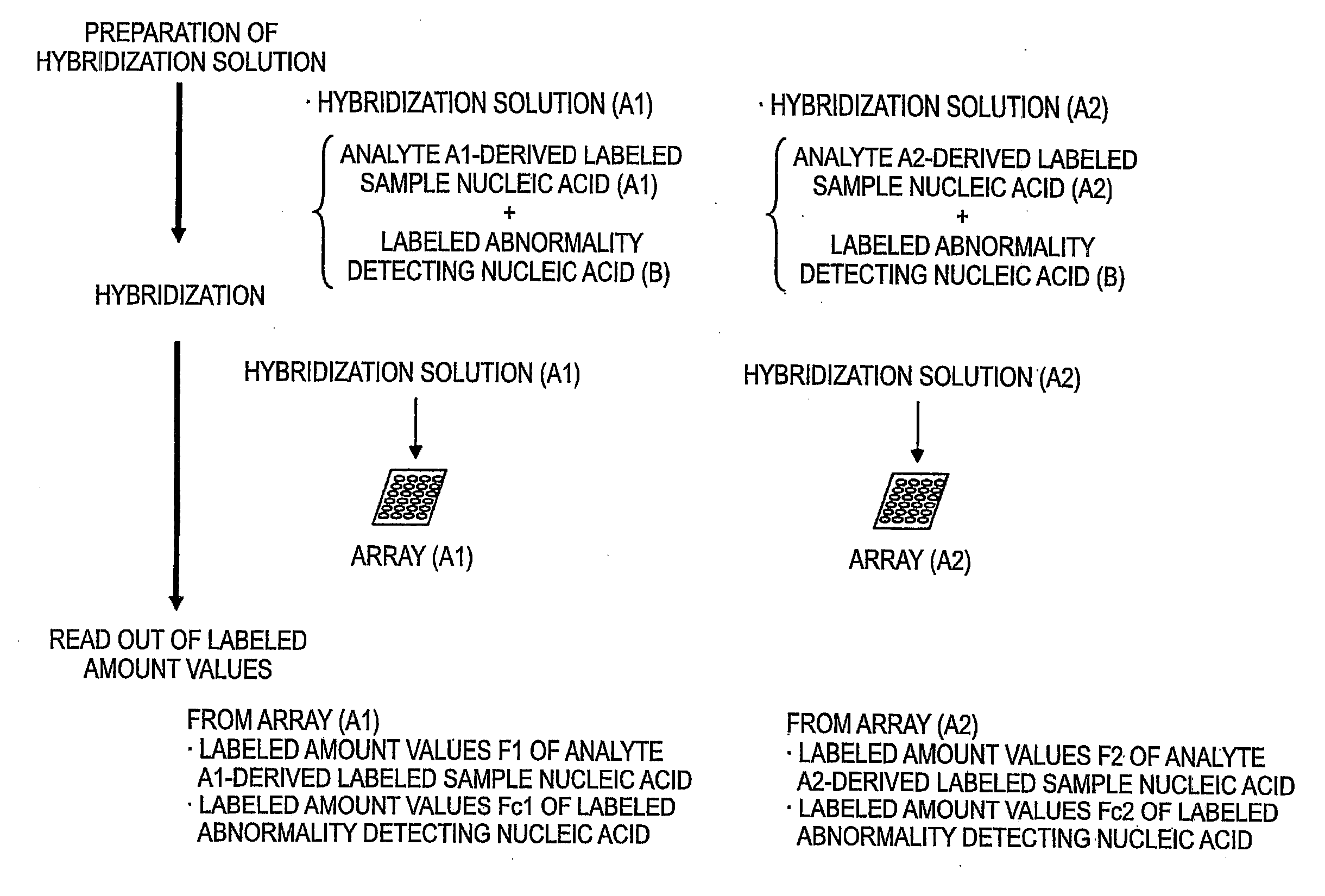
[0253]In this inventive example, it is assumed that a nucleic acid microarray 1 is possessed by the manufacturer and nucleic acid microarrays 2 and 3 are possessed by the user.

[0254]Cy3-Female DNA and Cy5-Male DNA were prepared by the same method of the of Comparative Example 1.

[0255]Cy5-Cot-1 DNA was prepared by the same method of the of Comparative Example 1. In this connection, all of the labeled abnormality detecting (correcting) nucleic acid Cy5-Cot-1 DNA samples to be hybridized with the nucleic acid microarrays 1 to 3 were used by subdividing from the same preparation batch.

[0256]This was prepared by the same method of the of Comparative Example 1.

[0257]This was prepared by the same method of the of Comparative Example 1. However, the hybridization solution containing labeled abnormality detecting (correcting) nucleic acid was prepared by adding and mixing, instead of the unpurif...

example 3

In the Case of Keeping at Least One Spot when all Spots of the Spots Spotted with the Same Probe were Determined as Abnormal Spots

[0275]Cy3-Sample A and Cy3-Sample B were prepared by the same method of the of Comparative Example 1. The sample A is DNA which was obtained from a cell which was obtained by immortalizing and cultivating patient specimen who is female and has defect in the chromosome 7. The sample B is DNA which was obtained from blood of a person who is female and has no genetic mutation.

[0276]Cy5-Cot-1 DNA was prepared by the same method of the of Comparative Example 1. In this connection, all of the labeled abnormality detecting (correcting) nucleic acid Cy5-Cot-1 DNA samples to be hybridized with the nucleic acid microarrays 1 and 2 were used by subdividing from the same preparation batch.

[0277]This was prepared by the same method of the of Comparative Example 1.

[0278]This was prepared by the same method of the of Comparative Example 1.

[0279]Pretreatment was carr...


PUM

PropertyMeasurementUnit
temperatureaaaaaaaaaa
timeaaaaaaaaaa

Abstract

A method carries out a nucleic acid analysis using a nucleic acid microarray. A probe nucleic acid including a probe sequence (a′) complementary to a target sequence (a) and a sequence (b′) which is different from the probe sequence (a′) is immobilized on the nucleic acid microarray. The method includes hybridizing the nucleic acid microarray and a labeled abnormality detecting nucleic acid (B) containing a sequence (b) which can be bound to the sequence (b′), obtaining a labeled amount value (Fc1) of the labeled abnormality detecting nucleic acid (B) from a spot (X1), and determining, based on the measured labeled amount value (Fc1), as to whether or not the spot (X1) is an abnormal spot unsuitable for detecting the target nucleic acid.

Description

CROSS-REFERENCE TO RELATED APPLICATIONS[0001]This application is based upon and claims the benefit of priority from the Japanese Patent Application No. 2009-128162 (filed on May 27, 2009), the entire contents of which are incorporated herein by reference.BACKGROUND[0002]1. Technical Field of the Invention[0003]The invention relates to a method for detecting abnormal spots, which are unsuitable for being used in evaluation of a hybridization result, among spots being present on a nucleic acid microarray by using an abnormality detecting nucleic acid, which can be hybridized with all spots, at the time of using a nucleic acid microarray.[0004]2. Description of the Related Art[0005]A comparative genomic hybridization (CGH) method is a technique which can detect abnormality of the number of genomic copies in the entire chromosome within a short period of time (A. Kallioniemi et al., Science, 258, 818-821 (1992)). However, this CGH method has a problem in detection resolution because it ...

Claims


Application Information

Patent Timeline
Patent Type & Authority Applications(United States)
IPC IPC(8): C40B30/04C40B40/06
CPCC12Q1/6837C12Q2525/161C12Q2545/101
Inventor YOSHIDA, JUNYAKANEHARA, HIDEYUKIUJIHARA, DAIINAZAWA, JOHJIIMOTO, ISSEI
Owner FUJIFILM CORP
Why Eureka
Try Eureka