
Endoscopic ultrasound-guided stent placement device and method

a technology of ultrasound guided stents and endoscopes, which is applied in the field of minimally invasive surgical methods, can solve the problems of inability to achieve video visualization (e.g., via a camera or other video element of an endoscope), inability to provide sufficient ultrasound reflectivity, and difficulty in navigating to a precise location within the body of the sten

Inactive Publication Date: 2010-12-16
WILSONCOOK MEDICAL +1
View PDF16 Cites 10 Cited by
  • Summary
  • Abstract
  • Description
  • Claims
  • Application Information

AI Technical Summary

Problems solved by technology

This is a particular challenge when a target region needs to be identified with sufficient locational specificity that visualization is preferable, but video visualization (e.g., via a camera or other video element of an endoscope) may be impractical.
However, these and polymer stents often do not provide sufficient difference in ultrasound reflectivity (relative to the surrounding tissues) to accurately direct and orient a stent under ultrasound visualization.
As a result, some prior art stents and similar access devices are difficult to navigate to a precise location within the body (the biliary tree being one example for illustrative purposes only).
This difficulty often occurs due to the lack of a guidance system configured to indicate where the device is located without introducing the risks associated with over-use of fluoroscopy.
For a variety of reasons, including the visualization challenges noted above, after the cannulator and / or dilator devices are withdrawn, it may be difficult to accurately navigate and position a stent in an optimal orientation.
If the procedure includes treatment of a cyst (for example, placement of a double-pigtail stent to treat a pseudocyst in the pancreas), a needle or cystotome may have to have been introduced and withdrawn (in which case, drainage / decompression of the cyst may further hamper introduction of a wire guide and stent in the manner typically used for stent placement).

Method used


Image

  • Endoscopic ultrasound-guided stent placement device and method
  • Endoscopic ultrasound-guided stent placement device and method
  • Endoscopic ultrasound-guided stent placement device and method

Examples


Embodiment Construction

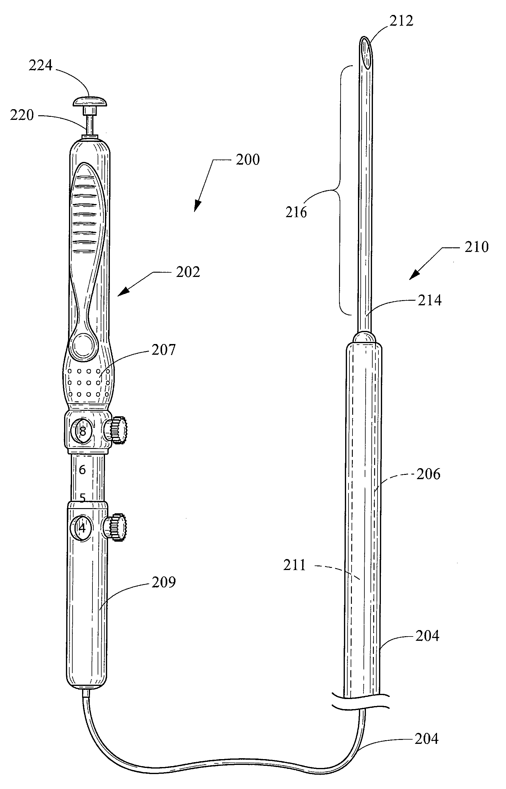
[0022]As used herein, including in the claims, the term “echogenic” is defined as having enhanced echogenicity. Specifically, it is used to refer to materials or portions of materials that are constructed or are treated to have greater reflectivity of ultrasonic waves than standard materials used for a stent, sheath, cannula, catheter, and / or stylet, and to provide an echogenic profile relative to surrounding tissues during use in a patient body to accurately orient and direct the echogenic device portion. It is known in the art that most materials used for a stent sheath, catheter, cannula, or stylet will reflect some ultrasonic waves, but the term “echogenicity,” as used herein includes treating the surface by creating a textured or patterned surface including, for example, one or more of dimples, divots, knurling, ridges, or the like—each of which is known in the art to enhance echogenicity as compared to a smooth surface for a similarly-sized / shaped object, (and / or, when specifi...


PUM


Abstract

A system and method for ultrasound-guided placement of a stent are provided. The system includes an echogenic cannula element that may include an echogenically-enhanced cannula that may be embodied as a piercing-tipped needle and / or an echogenically-enhanced stylet, where an echogenically-enhanced cannula element portion is near the distal cannula end. A stent is disposed about the echogenically-enhanced cannula element portion, such that the stent can be navigated to a target site using ultrasound imaging of the echogenically-enhanced cannula element portion.

Description

CROSS-REFERENCE TO RELATED APPLICATIONS[0001]This application claims priority to U.S. Provisional Application Ser. No. 61 / 186,593, filed Jun. 12, 2009, which is incorporated herein by reference in its entirety.TECHNICAL FIELD[0002]The invention relates generally to methods of minimally-invasive surgical methods. More particularly, the invention pertains to a system and method for ultrasound-guided placement of a stent.BACKGROUND[0003]The development of minimally invasive methods and devices over recent years has revolutionized the practice of medicine. These methods and devices allow clinicians to perform a wide variety of procedures while minimizing trauma to the patient. Along these lines, there is a need for devices and methods that employ minimally invasive technologies in order to access occluded regions in a mammalian body that may not be visible even from a minimally invasive device such as an endoscope. This is a particular challenge when a target region needs to be identifi...

Claims


Application Information

Patent Timeline
Patent Type & Authority Applications(United States)
IPC IPC(8): A61B8/12A61F2/84A61F2/04
CPCA61B2017/3413A61B2019/4857A61F2002/041A61F2/95A61B2019/5425A61B2090/0811A61B2090/3925
Inventor CLANCY, MICHAEL S.
Owner WILSONCOOK MEDICAL
Learn More