Gamma camera for performing nuclear mammography imaging

a nuclear mammography and camera technology, applied in the field of medical imaging systems, can solve the problems of increasing the patient's risk of not being able to detect a serious medical condition in time, affecting the sensitivity of the imaging, and affecting the patient's clinical experien

Active Publication Date: 2010-12-30
SMART BREAST CORP
View PDF12 Cites 22 Cited by
  • Summary
  • Abstract
  • Description
  • Claims
  • Application Information

AI Technical Summary

Problems solved by technology

This type of mammography imaging may not perform satisfactory imaging in women with dense breasts.
Moreover, some patients may experience discomfort due to the compression force applied to the breast and lower patient acceptance may result in missed examinations, thereby possibly increasing the patient's risk that a serious medical condition may not be detected in a timely fashion.
However, the size of these conventional detectors allows only anterior-posterior imaging with suffi

Method used


Image

  • Gamma camera for performing nuclear mammography imaging
  • Gamma camera for performing nuclear mammography imaging
  • Gamma camera for performing nuclear mammography imaging

Examples


Embodiment Construction

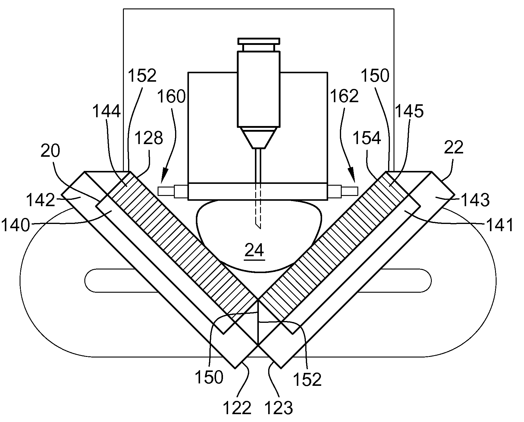
[0032]Various embodiments of the invention provide a system and method for performing molecular imaging of an anatomy of interest. A technical effect of the various embodiments is to provide a molecular imaging system that is configured to perform imaging optionally in both an H-mode and an L-mode configuration. The molecular imaging system is also configured to identify tumors or lesions during or after an imaging examination and to facilitate performing a biopsy of the identified tumors or lesions in the anatomy of interest.

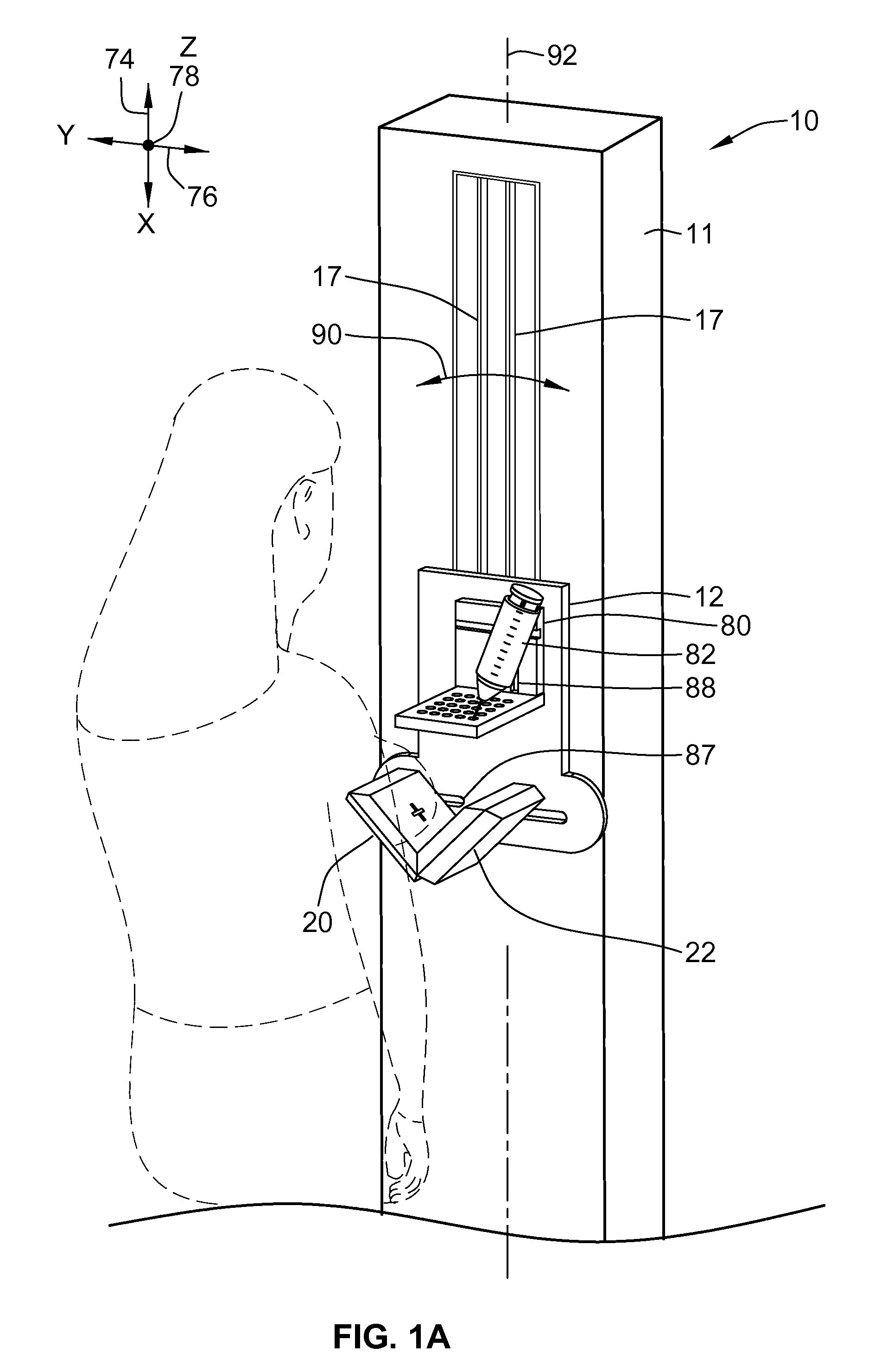
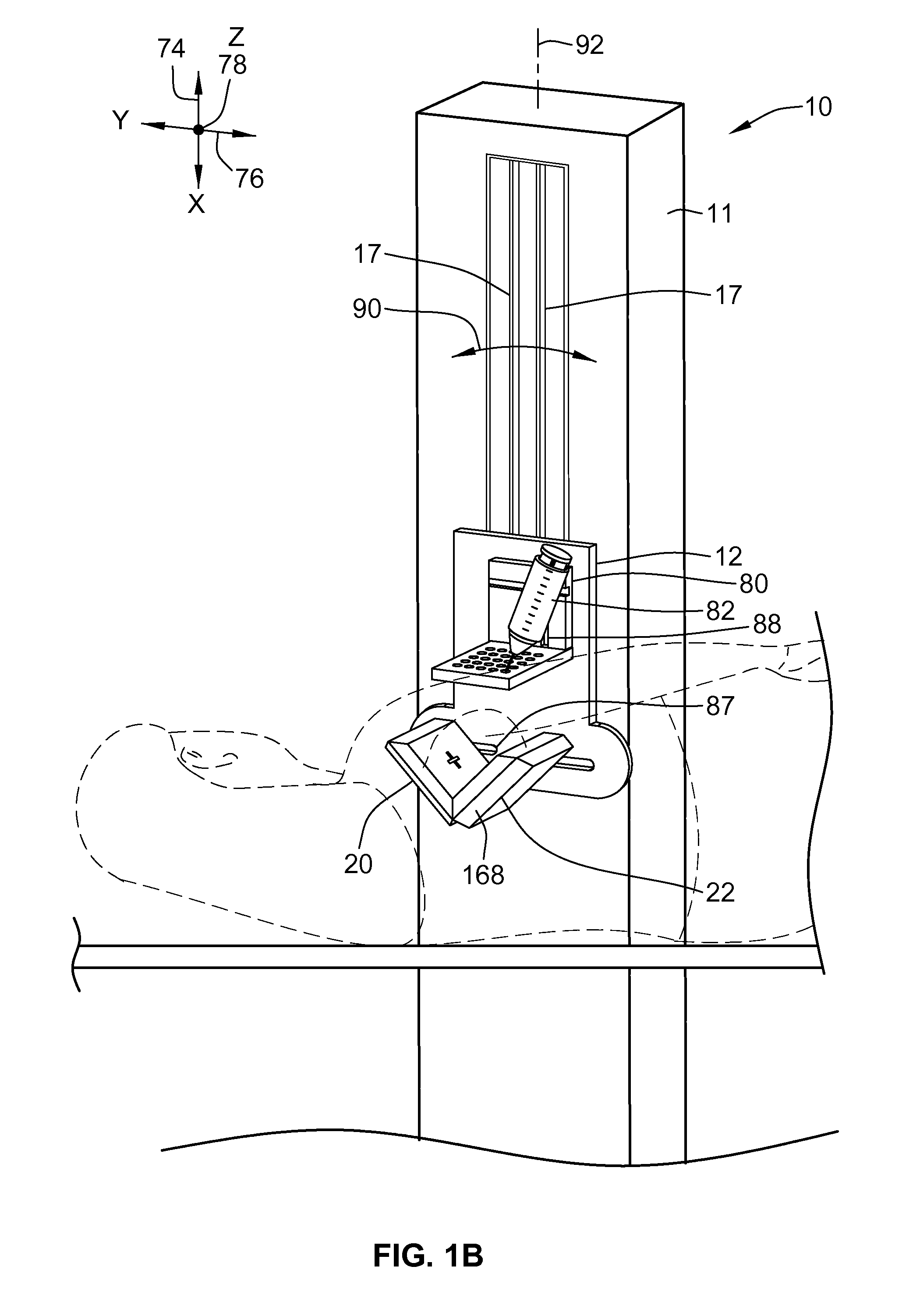
[0033]FIG. 1A is a front perspective view of an exemplary molecular imaging system 10 illustrating a patient positioned for imaging in a first imaging position. FIG. 1B is a front perspective view of the imaging system 10 illustrating a patient positioned for imaging in a second imaging position. FIG. 2 is a side view of the imaging system 10 shown in FIGS. 1A and 1B. In the exemplary embodiment, the molecular imaging system 10 is configured as a standalone mol...


PUM


Abstract

A gamma camera for use in a molecular imaging system includes a housing, a cadmium zinc telluride (CZT) camera disposed within the housing, and at least one retractable wall coupled to the housing and adapted to secure an anatomy of interest in a field-of-view of the gamma camera. The gamma camera also includes a chamfered edge to enable the gamma camera to contact a second gamma camera when the gamma camera is positioned in an L-mode imaging configuration.

Description

BACKGROUND OF THE INVENTION[0001]This invention relates generally to medical imaging systems, and more particularly to a system and method for performing nuclear mammography imaging.[0002]Different systems and methods for performing mammography imaging are known. For example, one conventional imaging system is a film screen mammography imaging. This type of mammography imaging system uses transmitted x-rays to produce an image of the breast. During the imaging procedure, a compression force is applied to a breast to improve image quality by reducing a thickness of the breast thereby spreading the breast tissue over a larger area. The reduction in the breast thickness, and spreading the breast over a larger area, facilitate reading of the projection radiographs, because the amount of “overlapping structures” within the imaged breast is minimized in the projection radiograph. This type of mammography imaging may not perform satisfactory imaging in women with dense breasts. Moreover, s...

Claims


Application Information

Patent Timeline
IPC IPC(8): A61B6/00G01T1/00
CPCA61B6/037Y10T29/49826A61B6/502A61B6/0414
Inventor BLEVIS, IRA
Owner SMART BREAST CORP
Why Eureka
Try Eureka