Multi-optical spectrum autofluorescence dislocation imaging reconstruction method based on single view

A technology of autofluorescence and tomography, applied in diagnosis, diagnostic recording/measurement, medical science, etc., can solve problems such as slow reconstruction speed, unfavorable real-time processing error, and inaccurate reconstruction light source

Inactive Publication Date: 2009-01-14
BEIJING UNIV OF TECH
View PDF0 Cites 30 Cited by
  • Summary
  • Abstract
  • Description
  • Claims
  • Application Information

AI Technical Summary

Problems solved by technology

[0004] The purpose of the present invention is to overcome the shortcomings of inaccurate reconstruction of light sources in autofluorescence tomography reconstruction methods, slow reconstruction speed, unfavorable real-time processing and possible errors in multispectral data acquisition, and proposes a multi

Method used


Image

  • Multi-optical spectrum autofluorescence dislocation imaging reconstruction method based on single view
  • Multi-optical spectrum autofluorescence dislocation imaging reconstruction method based on single view
  • Multi-optical spectrum autofluorescence dislocation imaging reconstruction method based on single view

Examples


Embodiment Construction

[0067] This embodiment will be described in detail below in conjunction with the accompanying drawings.

[0068] (1) Data acquisition:

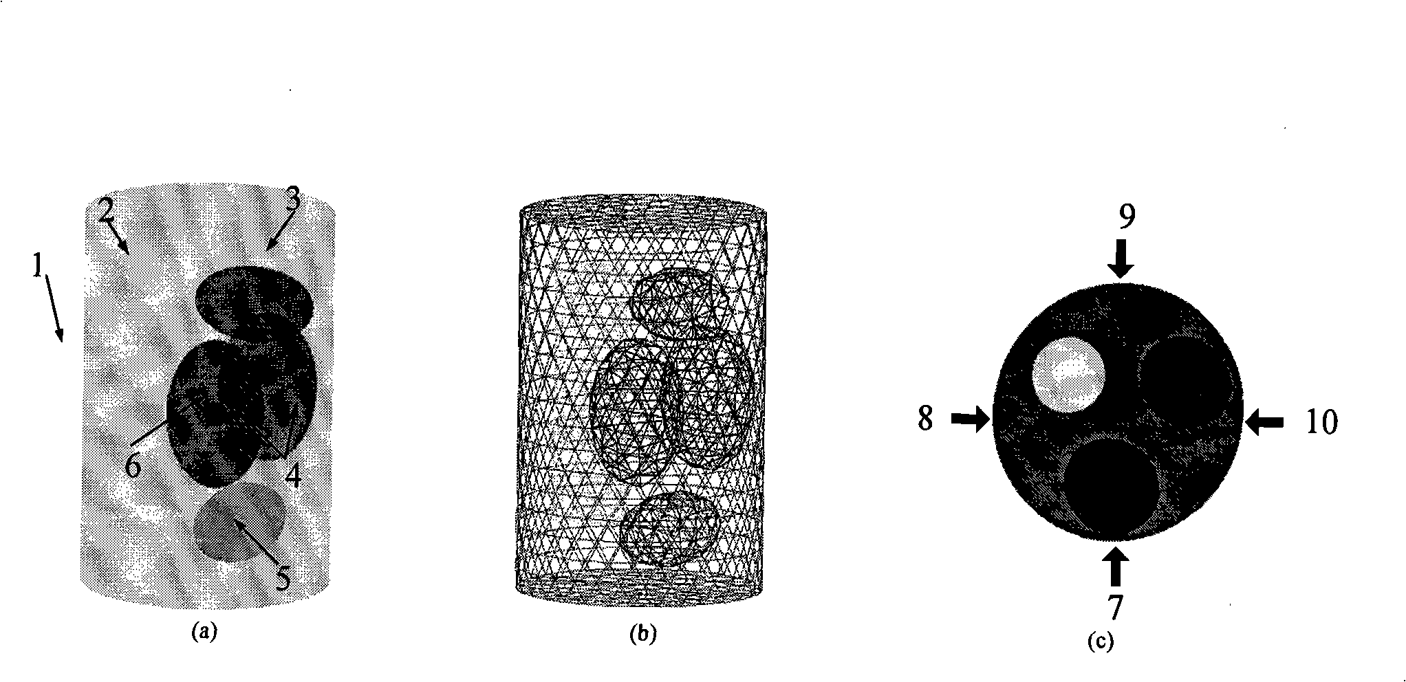
[0069] In order to verify this method, we designed a heterogeneous model to approximate the tissue in the mouse abdomen, such as figure 1 As shown in (a), the biological tissues are: muscle, bone, heart, lung, and liver, and the optical characteristic parameters of each tissue are shown in Table 1. In this experiment, we will reconstruct the light source density distribution of the spontaneous light source as the reconstruction target. In the reconstruction experiment, the size of the light source is 1 mm in radius, and the light source density is 0.238nano-Watts / mm 3 , the position is (-3, 5, 15). Its spectrum ranges from 500 nanometers to 750 nanometers. In order to conduct multi-spectral experiments, we divide this spectrum range into 5 spectral segments for separate detection based on the current detection level, respectively τ 1 =[500...


PUM


Abstract

The invention relates to a multi-spectrum auto fluorescence fault imaging reconstruction method based on single view, which belongs to the optical molecular imaging field. The method is based on the diffusivity equation model, considers the uneven characteristics of small animal body, and simultaneously also considers the spectrum and the practical application characteristics of the auto fluorescence light source. In order to achieve the purpose, the multi-spectrum auto fluorescence fault imaging reconstruction method based on single view comprises the flowing steps: firstly, data acquisition; secondly, the dispersing processing of finite element; thirdly, the optimization of the selection of the feasible light source zone; fourthly, the reconstruction of the light source. The method overcomes the defects that the reconstruction light source of the auto fluorescence fault imaging reconstruction method is inaccurate, the reconstruction speed is low, the real-time processing is not convenient, and error can exist during the multi-spectrum data acquisition.

Description

technical field [0001] The invention relates to an optical molecular imaging modality-autofluorescence tomography (BLT) technology, in particular to a single-view-based multispectral autofluorescence tomography reconstruction method. Background technique [0002] In the past few years, molecular imaging has received increasing attention due to its ability to reveal molecular and cellular information in vivo. Therefore, it has become an important tool for disease diagnosis and drug efficacy evaluation. As a small animal molecular imaging modality, optical imaging technology, especially autofluorescence tomography, has been rapidly developed and widely used because of its high performance, low price and non-invasive characteristics. Autofluorescence tomography is a recently developed optical imaging technique used to reconstruct the distribution of autofluorescent light sources in small living animals. Fluorescence occurs when luciferin and luciferase are exposed to oxygen a...

Claims


Application Information

Patent Timeline
IPC IPC(8): A61B5/00
Inventor 贾克斌冯金超田捷
Owner BEIJING UNIV OF TECH
Why Eureka
Try Eureka