Fluorescence focal modulation microscopy system and method

A microscopic system and focus technology, applied in the field of optical microscopy, can solve the problems of limited molecular imaging capabilities

Active Publication Date: 2010-08-11
NAT UNIV OF SINGAPORE
View PDF3 Cites 29 Cited by
  • Summary
  • Abstract
  • Description
  • Claims
  • Application Information

AI Technical Summary

Problems solved by technology

Although optical coherence tomography has been successfully applied to visualize fine structures in the human eye and hollow organs, its molecular imaging capabilities are rather limited

Method used


Image

  • Fluorescence focal modulation microscopy system and method
  • Fluorescence focal modulation microscopy system and method
  • Fluorescence focal modulation microscopy system and method

Examples


Embodiment Construction

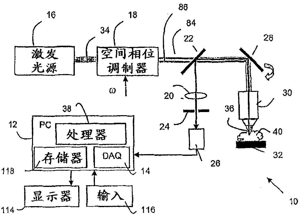
[0021] Focus modulation microscopy systems and methods are disclosed. Techniques according to embodiments of the present invention aim at imaging depths comparable to optical coherence tomography combined with molecular specificity. By using a spatial phase modulator in the excitation beam path, an intensity modulation is primarily achieved only in the focal volume, even when the focal point is located deep within the turbid medium. The oscillatory component in the detected fluorescence signal can be easily distinguished from the background signal caused by multiple scattering. Embodiments allow simultaneous acquisition of confocal microscopy images and focal modulation microscopy images. A series of image experiments were performed to demonstrate the advantages of focal modulation microscopy using tissue plant and cartilage tissue taken from chickens. Improved imaging penetration depth relative to conventional confocal microscopy systems can be achieved with focal modulatio...


PUM


Abstract

A fluorescence focal modulation microscopy system (10) and method (200) is disclosed for high resolution molecular imaging of thick biological tissues (40) with single photon excited fluorescence. Optical sectioning and diffraction limited spatial resolution are retained for imaging inside a multiple-scattering medium by the use of focal modulation, a technique for suppressing the background fluorescence signal excited by the scattered light. The focal modulation microscopy system has a spatial phase modulator (18) inserted in the excitation light path (34), which varies the spatial distribution of coherent excitation light around the focal volume periodically at a preset frequency. A fluorescence focal modulation image (122,142) is formed on a display (114) with the demodulated fluorescence, while a confocal image (120,140) is available simultaneously.

Description

technical field [0001] The present invention relates generally to optical microscopy, and more particularly to fluorescence confocal optical microscopy systems and methods. Background technique [0002] Various optical microscopes have been developed and used in modern biological research and clinical diagnosis. These light microscopes are often required to observe cells and their subcellular structures. The origin of modern cell biology begins with the discovery of cells more than 300 years ago as a direct result of the invention of the microscope. Conventional light microscopes have a very limited imaging depth. Only the surface microstructure can be observed, or the sample needs to be mechanically divided into very thin slices. The invention of confocal microscopy as described in US Pat. No. 3,013,467 led to a leap in imaging depth as a result of optical sectioning capability. Penetration depths of up to 200 microns can be achieved with biological tissue. Confocal mi...

Claims


Application Information

Patent Timeline
Patent Type & Authority Applications(China)
IPC IPC(8): G02B21/00
CPCG02B21/0076G02B21/0056G02B21/0032
Inventor NG·陈C·谢泼德C·H·翁
Owner NAT UNIV OF SINGAPORE
Why Eureka
Try Eureka