System for Processing Angiography and Ultrasound Image Data

a technology for processing systems and ultrasound catheters, applied in angiography, instruments, catheters, etc., can solve problems such as easy errors in the detection trajectory of the ultrasound catheter tip as it is slid backwards

Active Publication Date: 2011-02-10
SIEMENS HEALTHCARE GMBH
View PDF21 Cites 127 Cited by
  • Summary
  • Abstract
  • Description
  • Claims
  • Application Information

AI Technical Summary

Benefits of technology

[0006]A system corrects mis-alignment between a detected trajectory of an interventional ultrasound (IVUS) transducer during an IVUS retraction (pullback) procedure in which IVUS data is used in conjunction with angiographic X-ray data. A system provides a single composite image including multiple medical images of a portion of patient anatomy acquired using corresponding multiple different types of imaging device. A display processor generates data representing a single composite display image including, a first image area showing a first image of a portion of patient anatomy acquired using a first type of imaging device, a second image area showing a second imag

Problems solved by technology

The detected trajectory of the ultrasound catheter tip as it is slid backwards is prone to error which sho

Method used


Image

  • System for Processing Angiography and Ultrasound Image Data
  • System for Processing Angiography and Ultrasound Image Data
  • System for Processing Angiography and Ultrasound Image Data

Examples


Embodiment Construction

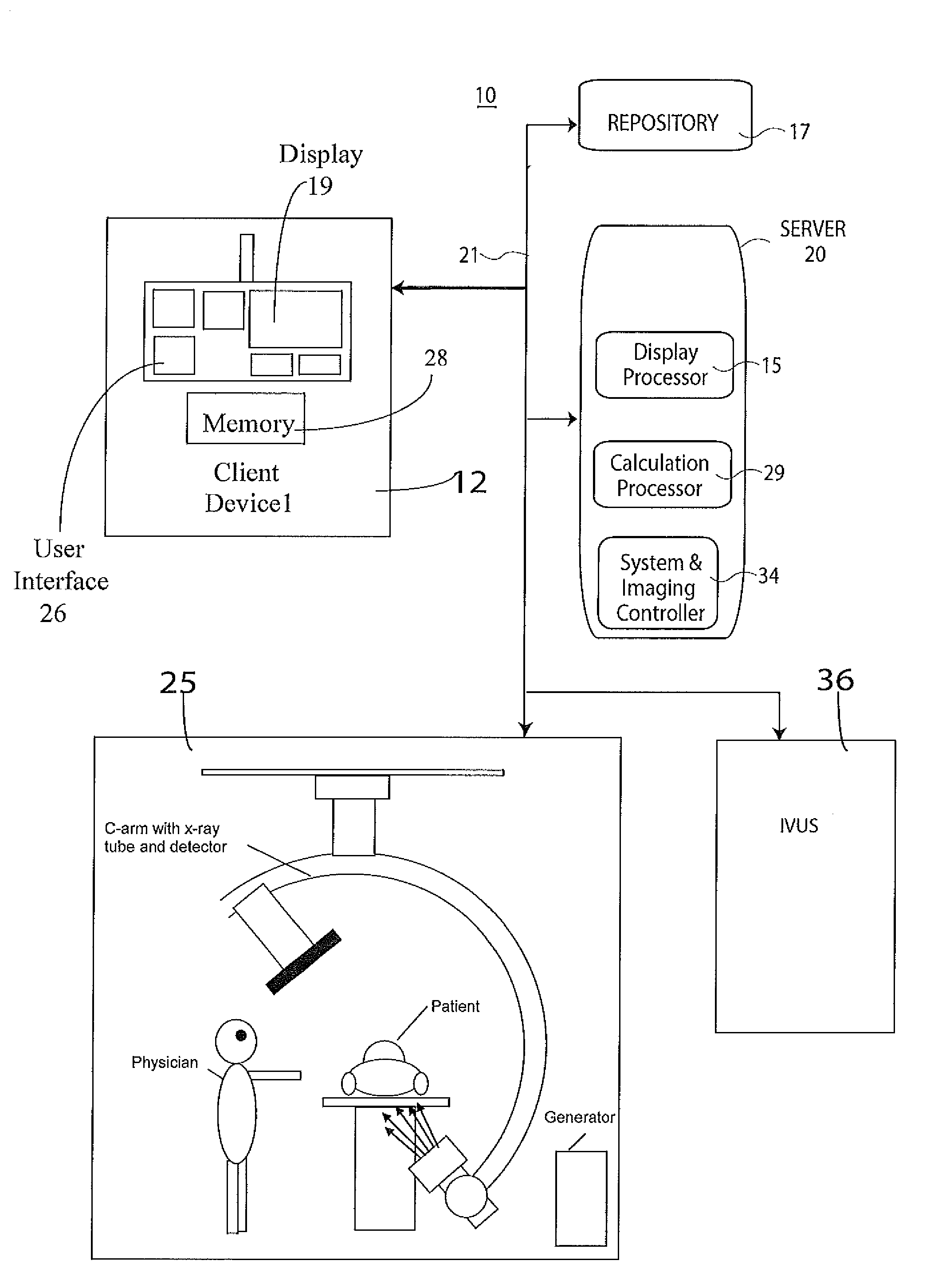
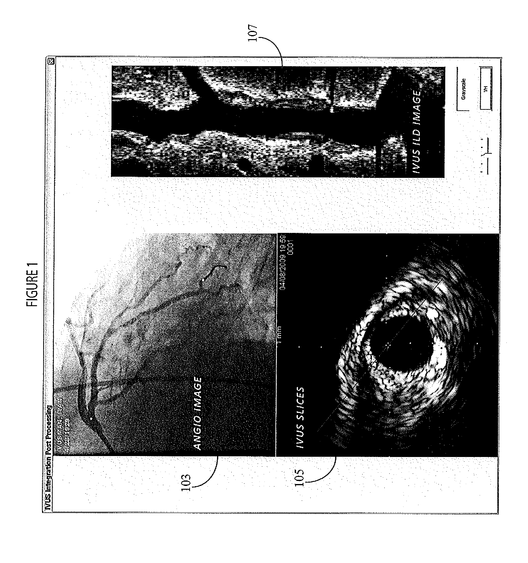
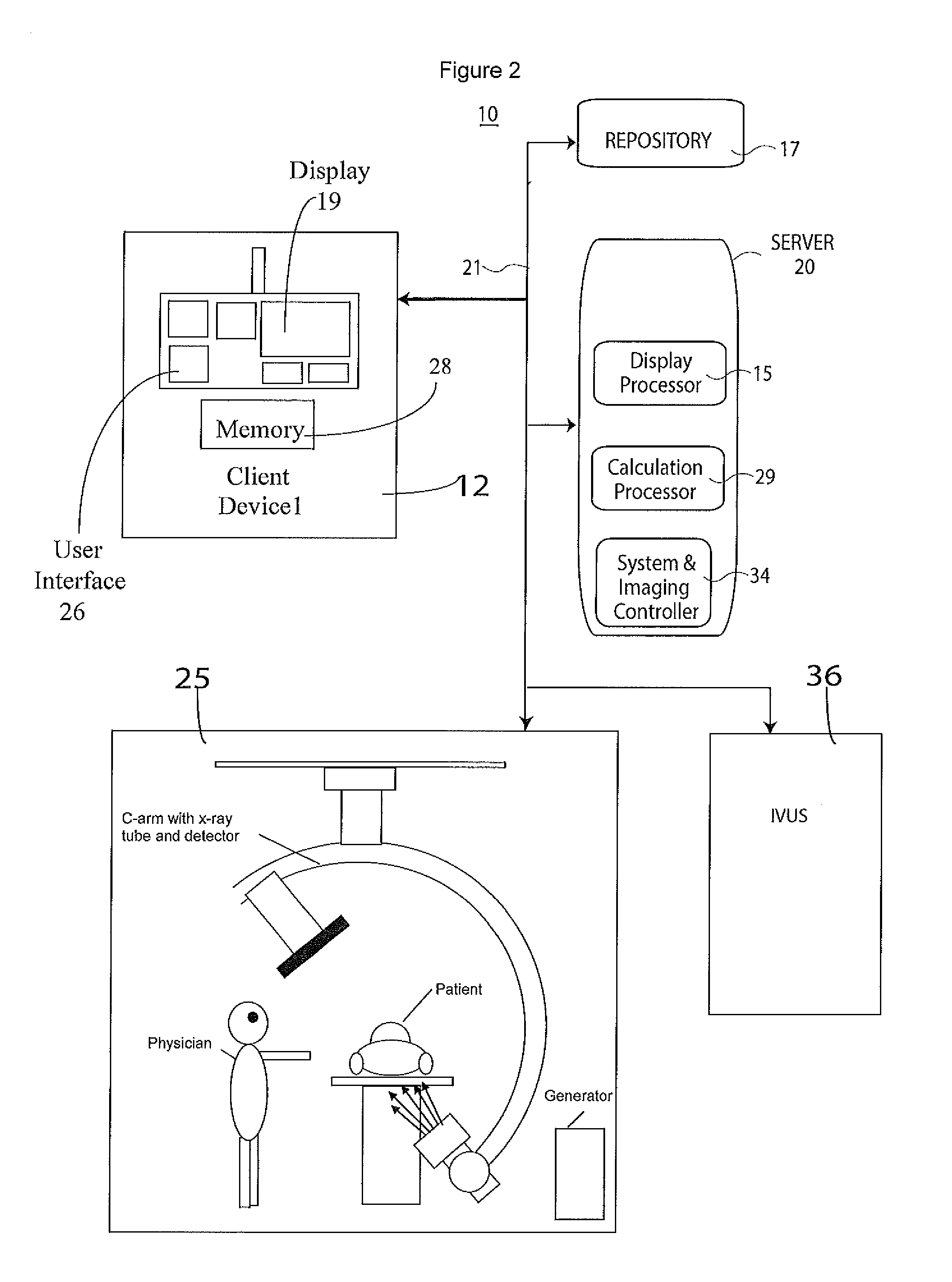
[0013]A system corrects mis-alignment (mis-registration) between a detected trajectory of an intravascular ultrasound (IVUS) transducer during an IVUS pullback procedure in which IVUS image data is used and aligned with angiographic X-ray data using timestamps and device localization to permit patient diagnosis. The system enables a user to correct a mis-registration between co-registered angiography and IVUS images. FIG. 1 illustrates a composite image showing angiography image 103 co-registered with IVUS image slice 105 presented together with an IVUS ILD (in-line display) pullback image 107 derived by retracting an IVUS catheter from a vessel. A user corrects mis-registration between co-registered angiography and IVUS images by selecting a point on either displayed image and moving it to a point that represents a correct registration with a marker present on the other image. A user may choose to correct either on the angiography image or on an IVUS image and the correction may be...


PUM


Abstract

A system provides a single composite image including multiple medical images of a portion of patient anatomy acquired using corresponding multiple different types of imaging device. A display processor generates data representing a single composite display image including, a first image area showing a first image of a portion of patient anatomy acquired using a first type of imaging device, a second image area showing a second image of the portion of patient anatomy acquired using a second type of imaging device different to the first type. The first and second image areas include first and second markers respectively. The first and second markers identify an estimated location of the same corresponding anatomical position in the portion of patient anatomy in the first and second images respectively. A user interface enables a user to move at least one of, (a) the first marker in the first image and (b) the second marker in the second image, to correct the estimated location so the first and second markers identify the same corresponding anatomical position in the portion of patient anatomy.

Description

[0001]This is a non-provisional application of provisional application Ser. No. 61 / 231,717 filed 6 Aug. 2009, by J. Baumgart.FIELD OF THE INVENTION[0002]This invention concerns a system for providing a single composite image including multiple medical images of a portion of patient anatomy acquired using corresponding multiple different types of imaging device.BACKGROUND OF THE INVENTION[0003]Angiography imaging modality and intravascular ultrasound (IVUS) imaging modality devices are both used to assess condition of patient blood vessels and each type of device has respective features and advantages. Known systems that use both these types of imaging modality device concurrently require that a location of an IVUS transducer be detected in live angiography images acquired during retraction (pullback) of an IVUS device from a vessel, for example. An intravascular ultrasound (IVUS) medical imaging modality device uses a catheter with a miniaturized ultrasound probe attached to a dista...

Claims


Application Information

Patent Timeline
IPC IPC(8): A61B5/055G09G5/00
CPCA61B5/489A61B6/4441A61B6/463A61B6/504A61B8/5238A61B8/12A61B8/4416A61B8/463A61B6/5247
Inventor BAUMGART, JOHN
Owner SIEMENS HEALTHCARE GMBH
Why Eureka
Try Eureka