
Method of manufacturing biosensor and biosensor produced thereby

a biosensor and biosensor technology, applied in the field of biosensors, can solve the problems of insufficient simplicity and rapidity of complicated operations, inability to measure without, and excessive sample amount, so as to reduce the required amount of liquid specimens, analyze the effect of test substances easily and quickly

Inactive Publication Date: 2011-02-24
PANASONIC CORP
View PDF2 Cites 9 Cited by
  • Summary
  • Abstract
  • Description
  • Claims
  • Application Information

AI Technical Summary

Benefits of technology

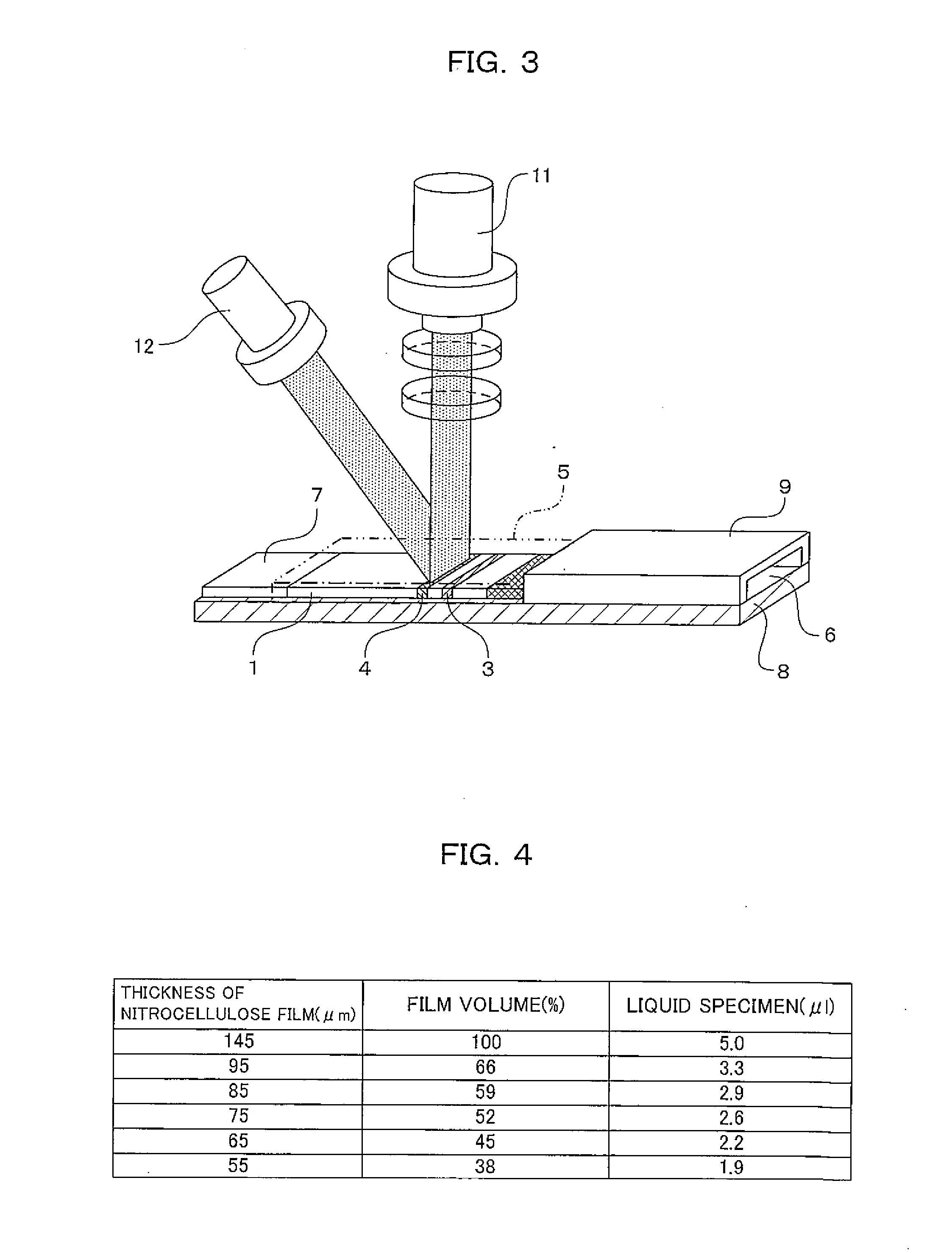
[0028]According to the present invention, in a method of manufacturing a biosensor in which a liquid specimen is developed to a development layer and a test substance in the liquid specimen is measured, a required amount of the liquid specimen can be reduced by reducing the thickness of the development layer and the development layer is 20 μm to 135 μm in thickness in the biosensor in which the liquid specimen is developed to the development layer and the test substance is measured in the liquid specimen. Thus it is possible to reduce the required amount of the liquid specimen and easily and quickly analyze the test substance from a trace amount of the liquid specimen with higher accuracy.

Problems solved by technology

Thus measurement cannot be conducted without supplying a large amount of liquid specimen.
Further, when the liquid specimen is added to the specimen adding portion, generally, blood cells that are formed elements are separated from plasma by using a centrifugal separator, and then a specified amount of the liquid specimen has to be added with an instrument such as a dispenser and a pipette, so that an excessive sample amount is necessary.
In a method of reducing an amount of liquid specimen (a required amount of liquid specimen) required for measurement, a small amount of liquid specimen diluted with a large amount of diluent is added, resulting in complicated operations with insufficient simplicity and rapidity.
In another method, a small amount of liquid specimen is added and then a large amount of developing solution is added to the liquid specimen, resulting in low simplicity and rapidity.
However, a reduction in the widths and lengths of the components of the biosensor makes it difficult to produce the biosensor.
For example, a reagent immobilizing portion to be measured is also reduced in size and manufacturing variations are likely to occur, so that the accuracy of measurement may decline.
However, even in the foregoing method, a reduction in the amount of liquid specimen is limited, resulting in limited response to market demand for reducing a burden on a patient by reducing the amount of liquid specimen.

Method used


Image

  • Method of manufacturing biosensor and biosensor produced thereby
  • Method of manufacturing biosensor and biosensor produced thereby
  • Method of manufacturing biosensor and biosensor produced thereby

Examples


example 1

The Quantification of Whole Blood CRP when a Development Layer is Reduced in Thickness

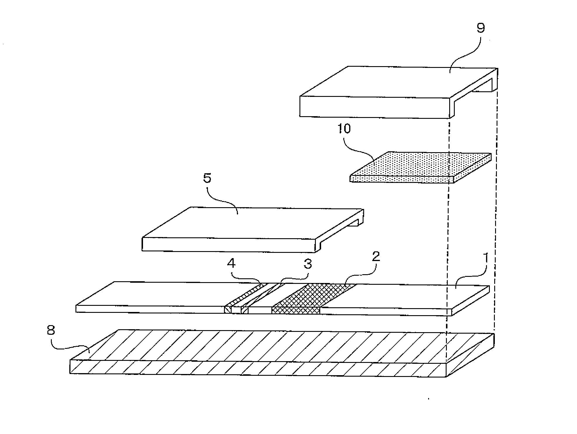
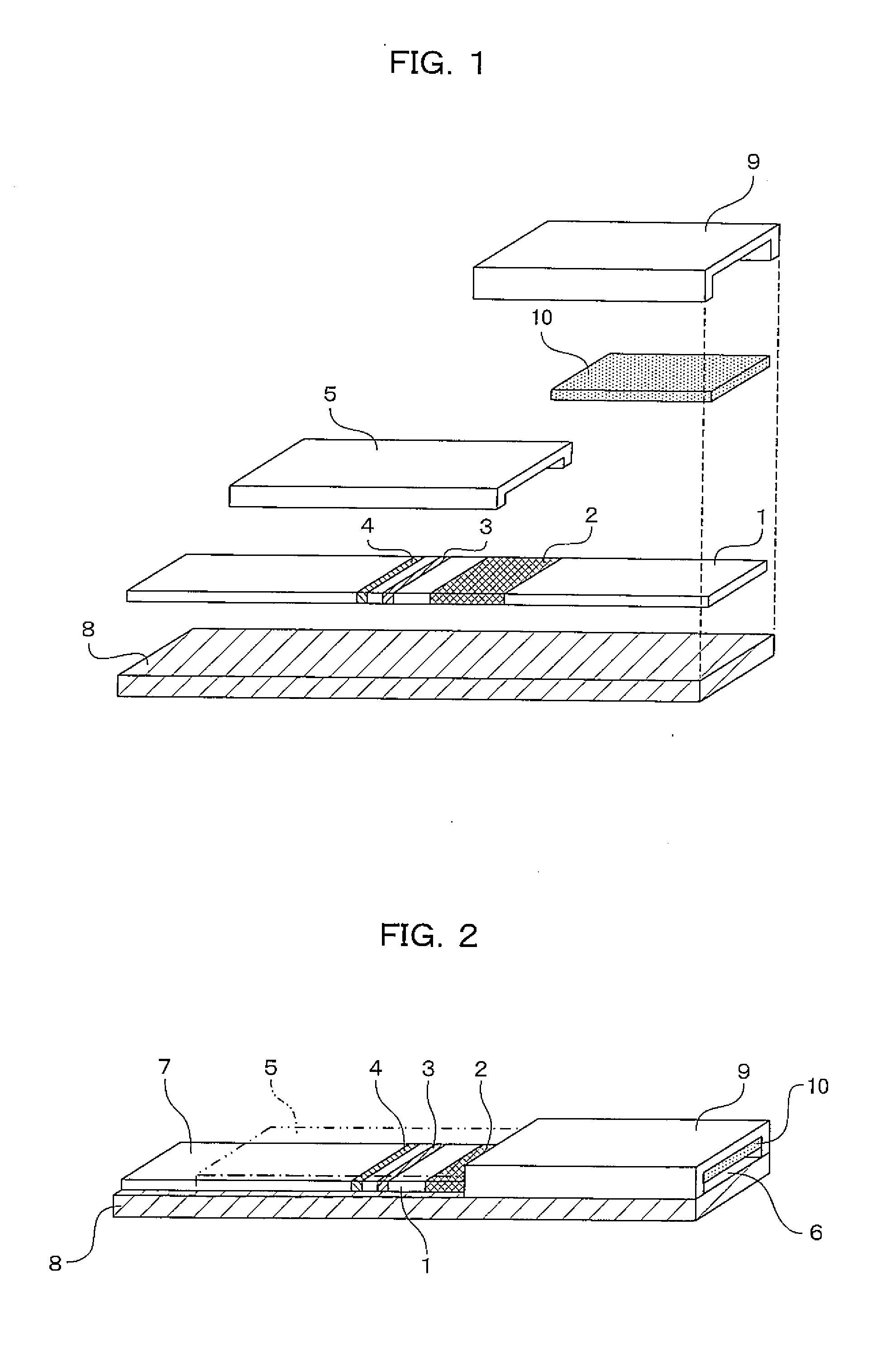
[0065]An immuno-chromatographic sensor was produced that was a biosensor including a first reagent immobilizing portion 3 in which anti-CRP antibody A was immobilized, a second reagent immobilizing portion 4 in which anti-CRP antibody B was immobilized, and a labeled reagent portion 2 containing a complex (labeled reagent) of anti-CRP antibody C and a gold colloid, on a development layer 1 including a nitrocellulose film. The immuno-chromatographic sensor is shown in FIGS. 1 and 2. In FIGS. 1 and 2, the immuno-chromatographic sensor includes the first reagent immobilizing portion 3 and the second reagent immobilizing portion 4 in which the antibodies are immobilized, the labeled reagent portion 2 that is provided between the reagent immobilizing portions and the adding portion of a liquid specimen and contains a complex of anti-CRP antibody C and a gold colloid, and a specimen adding portion 6.

[006...

example 2

The Quantification of Whole Blood CRP when a Development Layer is Reduced in Thickness

[0078]An immuno-chromatographic sensor was produced that was a biosensor including a first reagent immobilizing portion 3 in which anti-CRP antibody A was immobilized, a second reagent immobilizing portion 4 in which anti-CRP antibody B was immobilized, and a labeled reagent portion 2 containing a complex (labeled reagent) of anti-CRP antibody C and a gold colloid, on a development layer 1 including a nitrocellulose film. The immuno-chromatographic sensor is shown in FIGS. 1 and 2. In FIGS. 1 and 2, the immuno-chromatographic sensor includes the first reagent immobilizing portion 3 and the second reagent immobilizing portion 4 in which the antibodies are immobilized, the labeled reagent portion 2 that is provided between the reagent immobilizing portions and the adding portion of a liquid specimen and contains a complex of anti-CRP antibody C and a gold colloid, and a specimen adding portion 6.

[007...

example 3

The Quantification of Whole Blood CRP when the Thickness and the Pore Size of a Development Layer are Changed

[0084]An immuno-chromatographic sensor was produced that was a biosensor including a first reagent immobilizing portion 3 in which anti-CRP antibody A was immobilized, a second reagent immobilizing portion 4 in which anti-CRP antibody B was immobilized, and a labeled reagent portion 2 containing a complex (labeled reagent) of anti-CRP antibody C and a gold colloid, on a development layer 1 including a nitrocellulose film. The immuno-chromatographic sensor is shown in FIGS. 1 and 2. In FIGS. 1 and 2, the immuno-chromatographic sensor includes the first reagent immobilizing portion 3 and the second reagent immobilizing portion 4 in which the antibodies are immobilized, the labeled reagent portion 2 that is provided between the reagent immobilizing portions and the adding portion of a liquid specimen and contains a complex of anti-CRP antibody C and a gold colloid, and a specime...


PUM

PropertyMeasurementUnit
thicknessaaaaaaaaaa
thicknessaaaaaaaaaa
thicknessaaaaaaaaaa

Abstract

Provided are a biosensor and a method of manufacturing the same which can reduce an amount of liquid specimen required for measurement (a required amount of the liquid specimen), and conduct measurement with higher accuracy. In the biosensor, the liquid specimen is developed on a development layer by using chromatography and a test substance in the liquid specimen is measured. By reducing the thickness of the development layer (to 20 μm to 135 μm), the required amount of the liquid specimen is reduced. The thickness of the development layer is controlled to a smaller thickness thus, so that the required amount of the liquid specimen can be reduced in response to the measurement of a test substance in each liquid specimen in a state in which the accuracy of analysis is properly kept.

Description

TECHNICAL FIELD[0001]The present invention relates to a biosensor in which a test substance in a liquid specimen is developed to a development layer and is measured thereon, and particularly relates to a biosensor and a method of manufacturing the same which can reduce an amount of liquid specimen required for measurement.BACKGROUND ART[0002]In recent years, home care and community health care in doctor's offices and clinics have improved and the number of early diagnoses and the number of urgent laboratory tests have increased. Against this backdrop, analyzing devices have been demanded that can quickly and easily perform measurements with high accuracy even if users are not medical technologists. Thus small analyzing devices for POCT (Point of Care Testing) have received attention that can perform reliable measurements in a short time without complicated operations.[0003]POCT is a generic name for inspections conducted in locations “close to patients”, for example, in consulting r...

Claims


Application Information

Patent Timeline
Patent Type & Authority Applications(United States)
IPC IPC(8): C12M1/34
CPCG01N33/558G01N33/54388
Inventor KAWAMATA, RYOKOTAKAHASHI, MIE
Owner PANASONIC CORP
Learn More