
Methods, apparatus and articles of manufacture to process cardiac images to detect heart motion abnormalities

a technology of cardiac images and abnormal heart motion, applied in the field of cardiac images, can solve the problems of difficult classification and/or discrimination of heart motion based on distribution moments such as the mean systolic velocity, and difficulty in early detection of heart motion abnormalities, so as to reduce the time required for subjective evaluation of heart images, reduce inter and/or intra-observer variability, and reduce the effect of time required

Active Publication Date: 2011-03-17
GENERAL ELECTRIC CO +1
View PDF19 Cites 2 Cited by
  • Summary
  • Abstract
  • Description
  • Claims
  • Application Information

AI Technical Summary

Benefits of technology

[0005]The example methods, apparatus and articles of manufacture disclosed herein provide certain advantages over existing heart motion classification methods. For example, the subjective evaluation of heart images by a radiologist can be reduced and / or eliminated, thereby reducing inter and / or intra-observer variability. The examples disclosed herein also enabled automated analysis, which can reduce the time required to obtain a diagnosis and / or begin treatment.
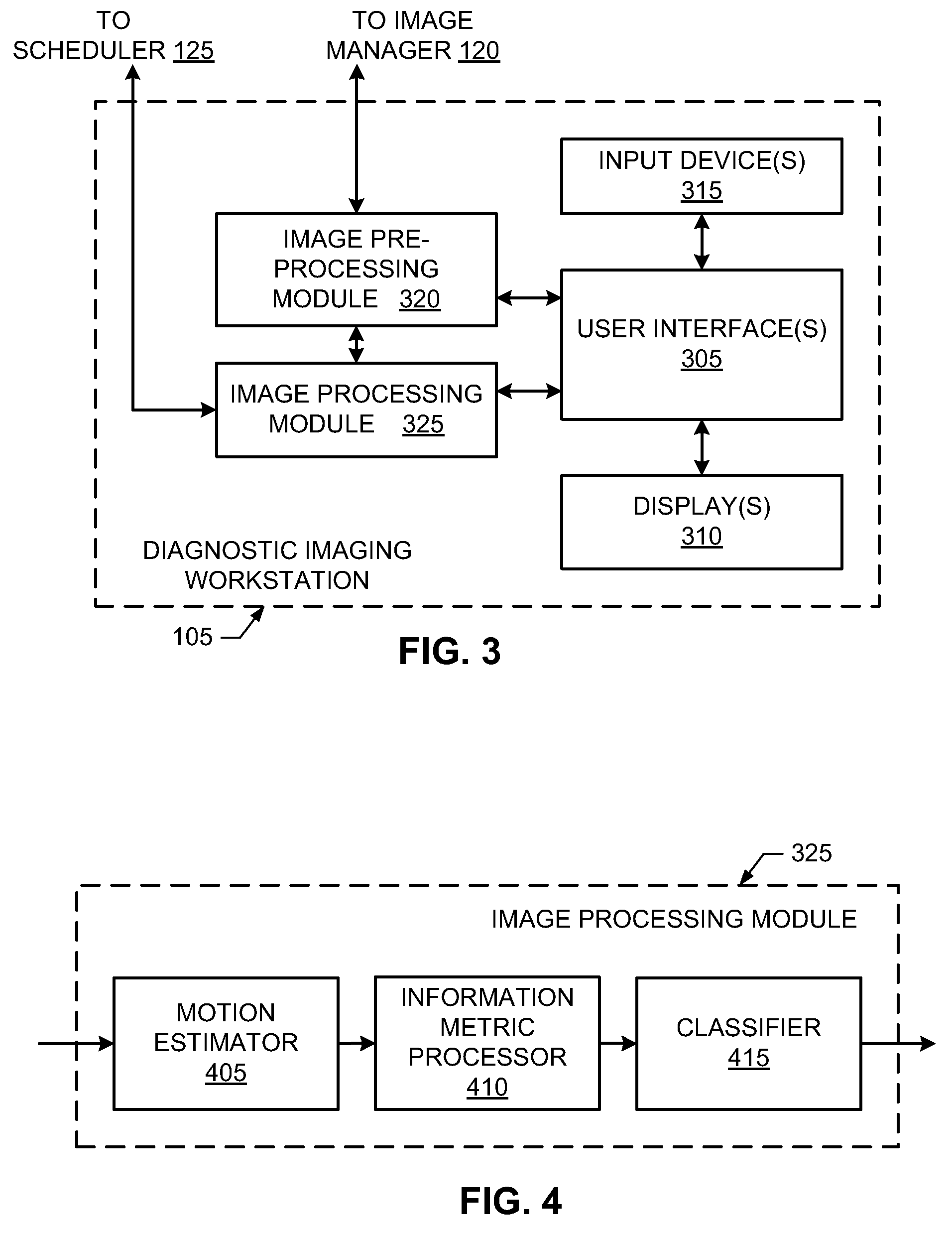
[0006]As disclosed herein, left-ventricle heart motion abnormalities may be detected by processing a sequence of cardiac images. The cardiac images may be segmented into one or more regions, and then processed or filtered with a cyclic model such as a recursive Bayesian filter or a Kalman filter. An example segmented region corresponds to the left-ventricular heart cavity. The cyclic model may be adapted and / or adjusted to apply temporal smoothing to the segmented cardiac images of the left-ventricular heart cavity. Such smoothing may be used to reduce the effect of image noise and / or segmentation inaccuracies. States and / or coefficients of the cyclic model may be used to characterize and / or represent the dynamics and / or motion of the segment left-ventricular heart cavity.

Problems solved by technology

However, due to the vast amount of information and uncertainty associated with heart motion, early detection of heart motion abnormalities may be difficult via visual inspection of cardiac images.
Due to statistical similarity between normal and abnormal hearts, the classification and / or discrimination of heart motion based on distribution moments such as the mean systolic velocity may be difficult and / or inaccurate.

Method used


Image

  • Methods, apparatus and articles of manufacture to process cardiac images to detect heart motion abnormalities
  • Methods, apparatus and articles of manufacture to process cardiac images to detect heart motion abnormalities
  • Methods, apparatus and articles of manufacture to process cardiac images to detect heart motion abnormalities

Examples


Embodiment Construction

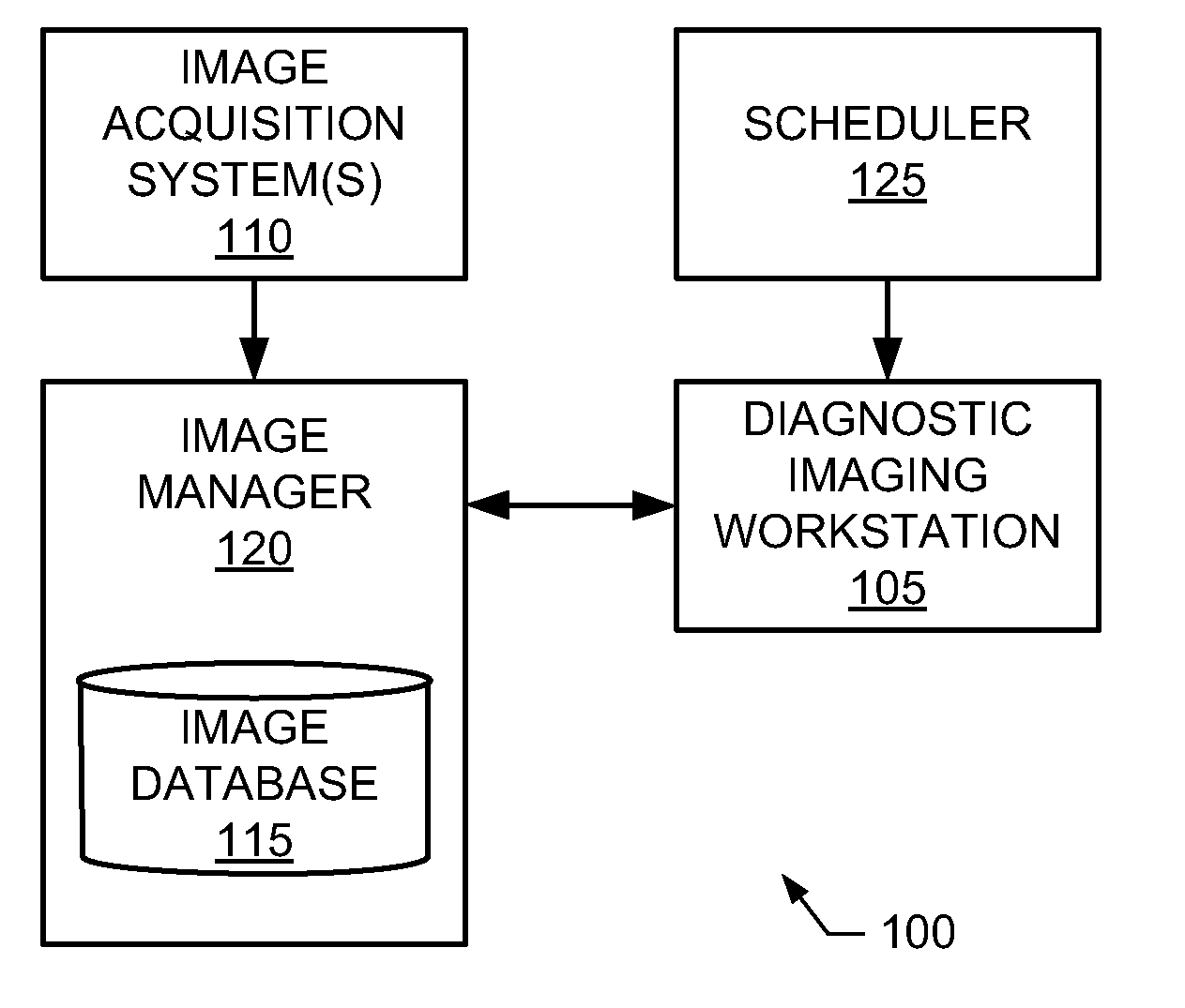
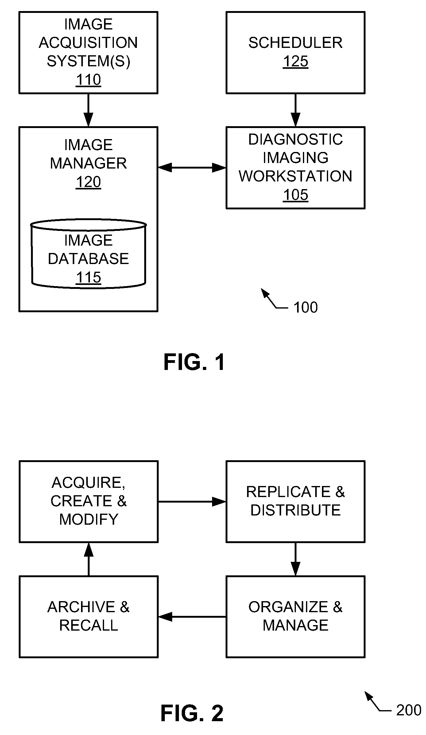
[0016]In the interest of brevity and clarity, throughout the following disclosure references will be made to an example diagnostic imaging workstation 105. However, the methods, apparatus and articles of manufacture described herein to process cardiac left-ventricle images to detect heart motion abnormalities may be implemented by and / or within any number and / or type(s) of additional and / or alternative diagnostic imaging systems. For example, the methods, apparatus and articles of manufacture described herein could be implemented by or within a device and / or system that captures diagnostic images (e.g., a computed tomography (CT) imaging system and / or magnetic resonance imaging (MRI) system), and / or by or within a system and / or workstation designed for use in viewing, analyzing, storing and / or archiving diagnostic images (e.g., the GE® picture archiving and communication system (PACS), and / or the GE advanced workstation (AW)). Moreover, the example methods, apparatus and articles of...


PUM


Abstract

Example methods, apparatus and articles of manufacture to process cardiac images to detect heart motion abnormalities are disclosed. A disclosed example method includes adapting a state of a state-space model based on a plurality of cardiac images to characterize motion of a heart, computing an information-theoretic metric from the state of the state-space model, and comparing the information-theoretic metric to a threshold to determine whether the motion of the heart is abnormal.

Description

RELATED APPLICATION[0001]This patents claims benefit from U.S. Provisional Patent Application Ser. No. 61 / 242,215, entitled “Methods, Apparatus and Articles of Manufacture to Detect Heart Motion Abnormalities,” and filed Sep. 14, 2009, which is hereby incorporated by reference in its entirety.FIELD OF THE DISCLOSURE[0002]This disclosure relates generally to cardiac images and, more particularly, to methods, apparatus and articles of manufacture to process cardiac images to detect heart motion abnormalities.BACKGROUND[0003]A widely used cardiac diagnostic technique involves the imaging of different portions of a heart during various phases of a heartbeat or cardiac cycle to detect or diagnose cardiac disease, abnormalities and / or damage. Example cardiac imaging tools are a magnetic resonance imaging (MRI) system and a computed topography (CT) imaging system.BRIEF DESCRIPTION OF THE INVENTION[0004]In view of the following descriptions and figures, it should be clear that the present d...

Claims


Application Information

Patent Timeline
IPC IPC(8): G06K9/62
CPCG06T2207/10081G06T2207/10088G06T2207/30048G06T7/0016G06T7/277G06V10/755G06V2201/03G06V10/761G06F18/22G06T7/0012
Inventor PUNITHAKUMAR, KUMARADEVANLI, SHUOBEN AYED, ISMAILROSS, IANISLAM, ALIRANKIN, RICHARD
Owner GENERAL ELECTRIC CO
Learn More