Method for acquiring x-ray data, x-ray device, computer program and electronically readable storage medium

a technology of x-ray data and x-ray device, which is applied in the field of acquiring x-ray data, x-ray device, computer program and electronically readable storage medium, can solve the problems of insufficient rate of 15 images per second to sample heart motion adequately, diametrically opposed to the trend of decreasing patient x-ray dose or keeping patient dose low, and reducing patient x-ray dose and contrast agent. , the effect of increasing the accuracy of overlaying/

Active Publication Date: 2018-12-27
SIEMENS HEALTHCARE GMBH
View PDF5 Cites 2 Cited by
  • Summary
  • Abstract
  • Description
  • Claims
  • Application Information

AI Technical Summary

Benefits of technology

[0015]Embodiments provide a method for more robustly and accurately evaluating x-ray images associated with heart cycle phases together.
[0022]Embodiments provide ECG-synchronous acquisition of x-ray data sets, increasing the accuracy and robustness in comparison to a non-ECG-synchronous acquisition technique.
[0024]In an embodiment, at least one of the at least one first x-ray image data set is a contrast-enhanced anatomy data set and at least one of the at least one second image data set includes intra-operational fluoroscopy images. The fluoroscopy images are displayed overlaid or fused with anatomy images of the first x-ray image data set including the same heart cycle phase. The first x-ray image data set depicts the anatomy and is used as a background for current fluoroscopy images depicting an instrument used in a minimally invasive intervention. A contrast-enhanced anatomy data set may also be called a “dynamic road map”. Embodiments provide for ECG-synchronous acquisition of the dynamic road map, that may be ECG-synchronously overlaid to the fluoroscopy images depicting the medical instrument used in the minimally-invasive intervention. The accuracy of overlaying / fusing the dynamic road map is considerably increased. Since the phases of the fluoroscopy images correspond to the phases of the contrast-enhanced anatomy data set by adequately controlling their acquisition anyway, it is sufficient to record the dynamic road map over one heart cycle with one contrast agent filling only as opposed to using multiple heart cycles for acquisition of a dynamic road map. Embodiments reduce patient x-ray dose and contrast agent. In other embodiments, image data sets are evaluated together, for example when comparing structures in these data sets or deriving other results.
[0025]The contrast-enhanced anatomy data set and the fluoroscopy images are acquired using the same x-ray device, for example, with the patient positioned for the operation to be performed. The first x-ray image data set and the second x-ray image data set are inherently registered to one another, facilitating the combined evaluation / overlay or fusion. The x-ray device may be an angiography device, for example, including a C-arm, to which the x-ray source and the x-ray detector are mounted at opposing ends.
[0027]In an embodiment, the contrast-enhanced anatomy data set may include digital subtraction angiography images (DSA images). Mask images and fill images are matched in heart cycle phase by acquiring the images triggered at the beginning of each heart cycle covered using an acquisition rate such that the phases of the mask images and the fill images match. It is thus possible to acquire mask images during at least one heart cycle natively, that is, without using contrast agent, and acquire fill images during at least one further heart cycle after contrast agent has been administered. Due to both acquisitions performed ECG-synchronized, the phases match such that the mask images may be subtracted from the fill images to create a dynamic DSA road map. The dynamic DSA road map facilitates blood vessel segmentation significantly since the detection algorithms for contrasts-filled blood vessels are not distracted by other native structures in the images.
[0032]The acquisition rate may be chosen dependent on user input. The user input may be, for example choosing a certain number of images per heart cycle to be acquired. However, the user may also choose a certain acquisition rate that may be adapted later on during the acquisition as described above. It is also possible that the user input is constrained, for example, if certain requirements are to be met. If, for example, the first x-ray image data set is a four-dimensional CT data set with a predetermined number of phases for which a volume image is included, the user may be confined to using integer divisors of the number of phases contained in the four-dimensional CT data set. Corresponding constraints may also result when using a dynamic route map acquired before. Reducing the number of phases imaged for the second x-ray image data set may also be advantageous to further reduce patient x-ray dose.

Problems solved by technology

However, the anatomical structures of interest are more or less transparent to x-rays, meaning the structures are not visible in fluoroscopy images without using contrast agents.
However, known static fusion and overlay techniques do not allow precise fusion and / or overlay of interesting structures regarding the heart region of the patient, since the region is subject to heart motion and breathing motion.
For example, when overlaying x-ray images, inaccuracies occur, since, among others, the motion of the blood vessel is too fast, so that usual acquisition rates of for example 15 images per second do not suffice to sample the heart motion adequately.
This approach, however, is diametrically opposed to the trend of decreasing patient x-ray dose or to keep patient dose low, respectively.

Method used


Image

  • Method for acquiring x-ray data, x-ray device, computer program and electronically readable storage medium
  • Method for acquiring x-ray data, x-ray device, computer program and electronically readable storage medium

Examples


Embodiment Construction

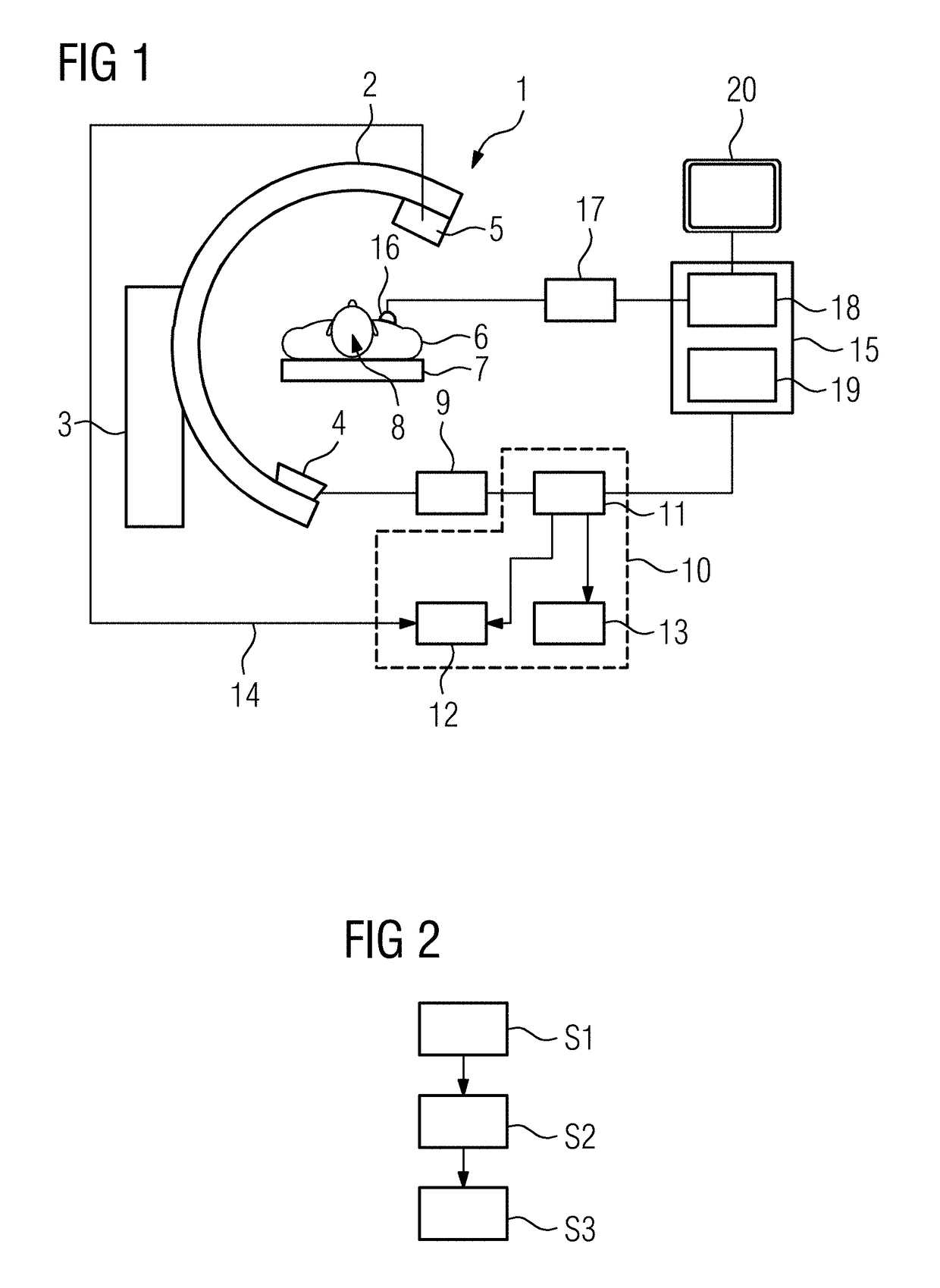
[0042]FIG. 1 depicts an example x-ray device 1, that is an angiography device including a C-arm 2 supported by a support 3. To opposite ends of the C-arm 2, an x-ray source 4 and an x-ray detector 5 are mounted. The C-arm may be positioned relatively to a patient 6 supported by a patient table 7 to realize different acquisition geometries.

[0043]The x-ray device 1 may, for example, be used to survey a minimally invasive intervention / operation of a patient 6, for example, for tracking a medical instrument 8 inserted into a patient 6, for example a catheter.

[0044]A voltage for the x-ray source 4, e.g. an x-ray tube, is provided by an x-ray generator 9 that receives control signals from control device 10, that is configured to perform the steps of a method. The control device 10 includes at least a control unit 11, an image evaluation unit 12 and a user interface 13. The image evaluation unit 12 also receives raw x-ray data from the x-ray detector 5 according to arrow 14.

[0045]The x-ray...


PUM


Abstract

A method and device for acquiring at least a part of an x-ray data set with at least one x-ray device. The x-ray data set includes at least one first x-ray image data set and at least one second x-ray image data set acquired separated in time that are to be evaluated together. Both x-ray image data sets show a region of interest of a patient that is subject to heart motion at different phases of a heart cycle. The second x-ray image data set is acquired triggered at a beginning of each heart cycle covered by the acquisition and with an acquisition rate such that the phases of the acquired x-ray image data set match phases of the first x-ray image data set.

Description

CROSS REFERENCE TO RELATED APPLICATIONS[0001]This application claims the benefit of EP17178141.2 filed on Jun. 27, 2017, which is hereby incorporated by reference in its entirety.FIELD[0002]Embodiments relate to a method for acquiring at least a part of an x-ray data set with at least one x-ray device, the x-ray data set including at least one first x-ray image data set and at least one second x-ray image data set acquired separated in time, that are to be evaluated together, wherein both x-ray image data sets show a region of interest of a patient that is subject to heart motion, at different phases of a heart cycle.BACKGROUND[0003]Heart diseases close to the human heart, for example coronary artery chronic total occlusion (CTO), are one of the leading causes of death of patients. Thus, minimally invasive operation techniques have been developed to treat patients suffering from heart diseases. For example, to treat CTO, an available procedure is percutaneous coronary intervention (...

Claims


Application Information

Patent Timeline
Patent Type & Authority Applications(United States)
IPC IPC(8): A61B6/00G06T7/00A61B5/352
CPCA61B6/5205G06T7/0016A61B6/504A61B6/5235A61B6/5264A61B5/7285A61B6/032A61B6/12A61B6/405A61B6/463A61B6/466A61B6/481A61B6/486A61B6/487A61B6/503A61B6/5288A61B6/541A61B6/545A61B2576/023G16H30/40A61B2090/376A61B5/318A61B5/352
Inventor MEYER, ANDREAS
Owner SIEMENS HEALTHCARE GMBH
Why Eureka
Try Eureka