Method for detection and quantification of plk1 expression and activity
a technology of expression and activity, applied in the field of polo like kinase 1 (), can solve the problems of limited success in the effort to develop useful therapies to regulate the hyper-proliferative properties of cancer cells, and achieve the effect of reducing the number of attempts to develop useful therapies
- Summary
- Abstract
- Description
- Claims
- Application Information
AI Technical Summary
Problems solved by technology
Method used
Image
Examples
example 1
Construction of GST-PBIPtides
[0161]This example describes exemplary techniques to construct PBIPtides used in subsequent studies.
[0162]To generate GST-PBIPtide expressing constructs, a pUC19 derivative, pUC19N, was generated in which multiple cloning sites were restructured to contain both BamHI and BglII. A small DNA fragment encoding GGPGG (SEQ ID NO: 12) fused-YETFDPPLHSTAIYADEE (amino acid residues 68-85 of SEQ ID NO: 10) (PBIPtide) was digested with BamHI (5′ end) and BglII (3′ end) and then inserted into pUC19N digested with the corresponding enzymes. The GGPGG (SEQ ID NO: 12) sequence was added at the N terminus of the PBIPtide as a linker between PBIPtide repeats. The resulting pUC19NGGPGG-PBIPtide was digested with BglII and then ligated with another copy of GGPGG-PBIPtide digested with BamHI (5′ end) and BglII (3′ end), yielding pUC19N-GGPGG-PBIPtide2. These cloning steps were repeated 2 more times to generate pUC19N-GGPGG-PBIPtide4 (this cloning strategy disables the N-te...
example 2
Exemplary Plk1 Kinase Assay Using GST-PBIPtide as a Plk1 Affinity Ligand and in vitro Substrate
[0164]This example describes tests for Plk1 kinase activity using a GST-PBIPtide as a Plk1 affinity ligand and in vitro substrate of Plk1.
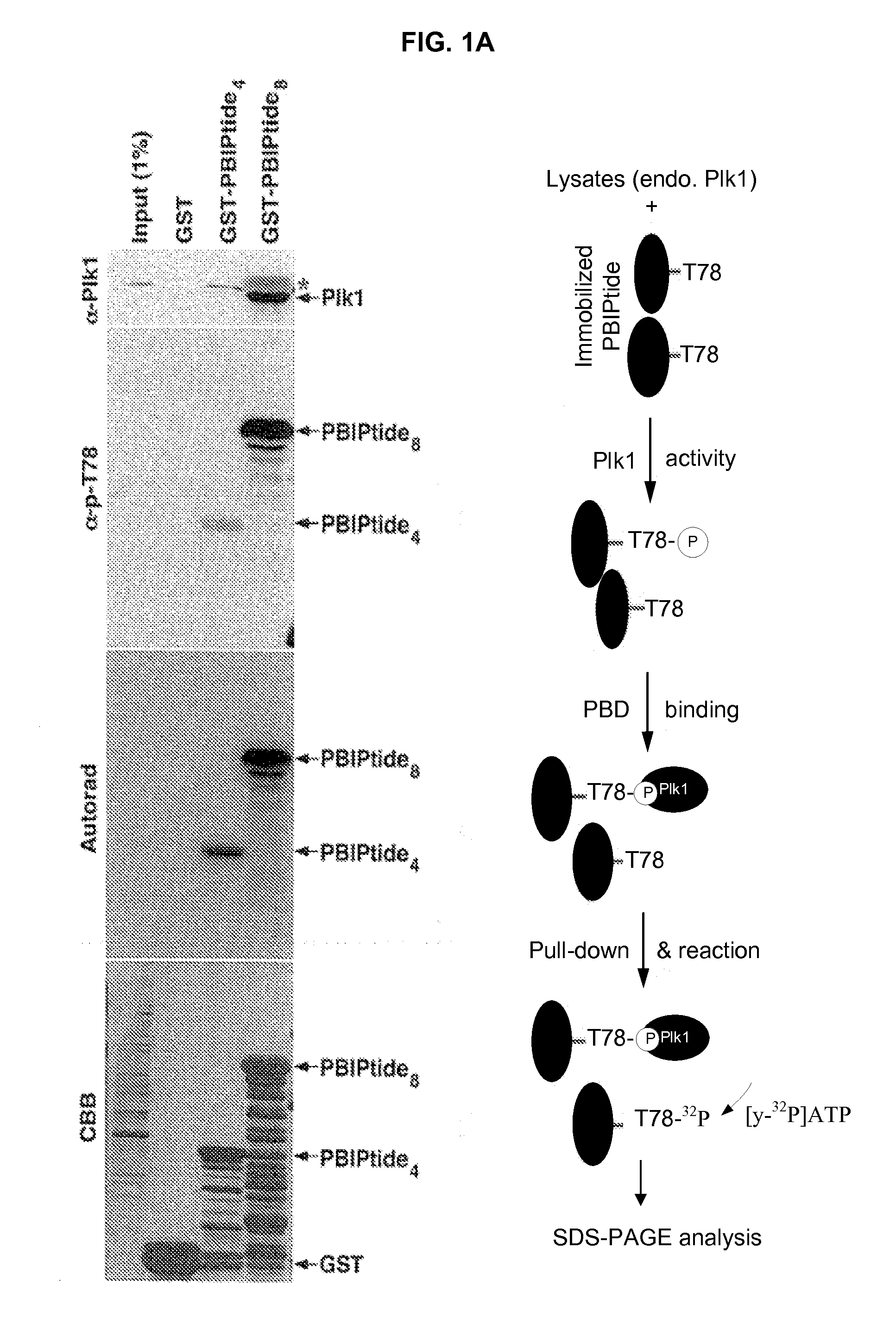
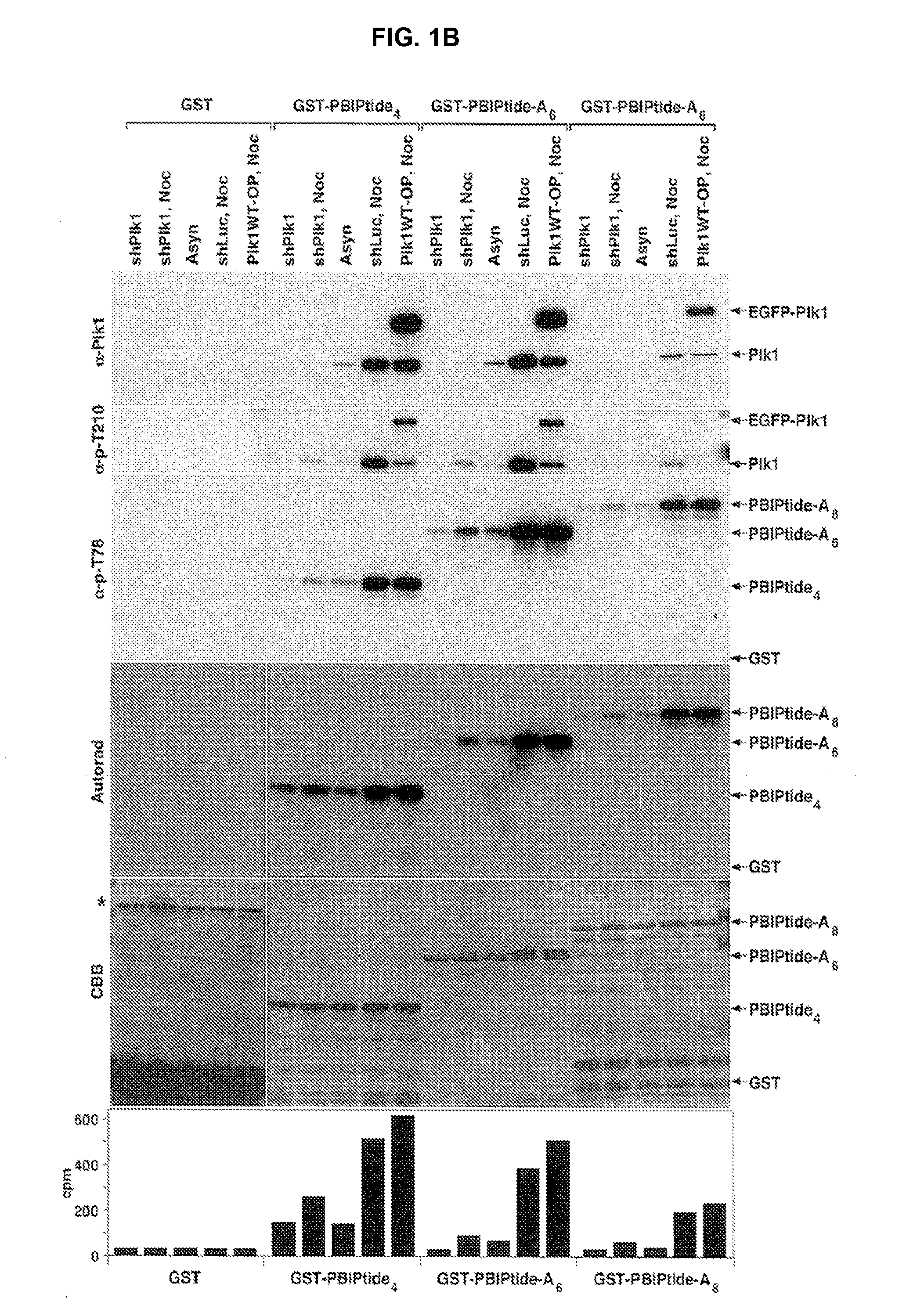
[0165]Plk1 efficiently phosphorylates a centromeric protein PBIP1 at T78, and this phosphorylation event generates a docking site for a high-affinity interaction between the PBD of Plk1 and p-T78 PBIP1 (Kang et al. (2006) Mol Cell 24:409-422). Subsequent investigation revealed that none of the other mitotic kinases tested (Cdc2, Aurora A, Aurora B, Mps1, and Erk1) appeared to phosphorylate the T78 residue of PBIP1. By taking advantage of the specific Plk1-dependent PBIP1 phosphorylation and subsequent interaction between the resulting p-T78 epitope and the PBD, it was examined whether a GST-fused PBIP1 peptide bearing the T78 motif (hereon referred to as GST-PBIPtide) could precipitate Plk1 through the Plk1-generated p-T78 epitope and whether the precipi...
example 3
Specificity of PBIPtides for Plk1
[0174]This example describes tests for the specificity of the GST-PBIPtides disclosed herein for Plk1.
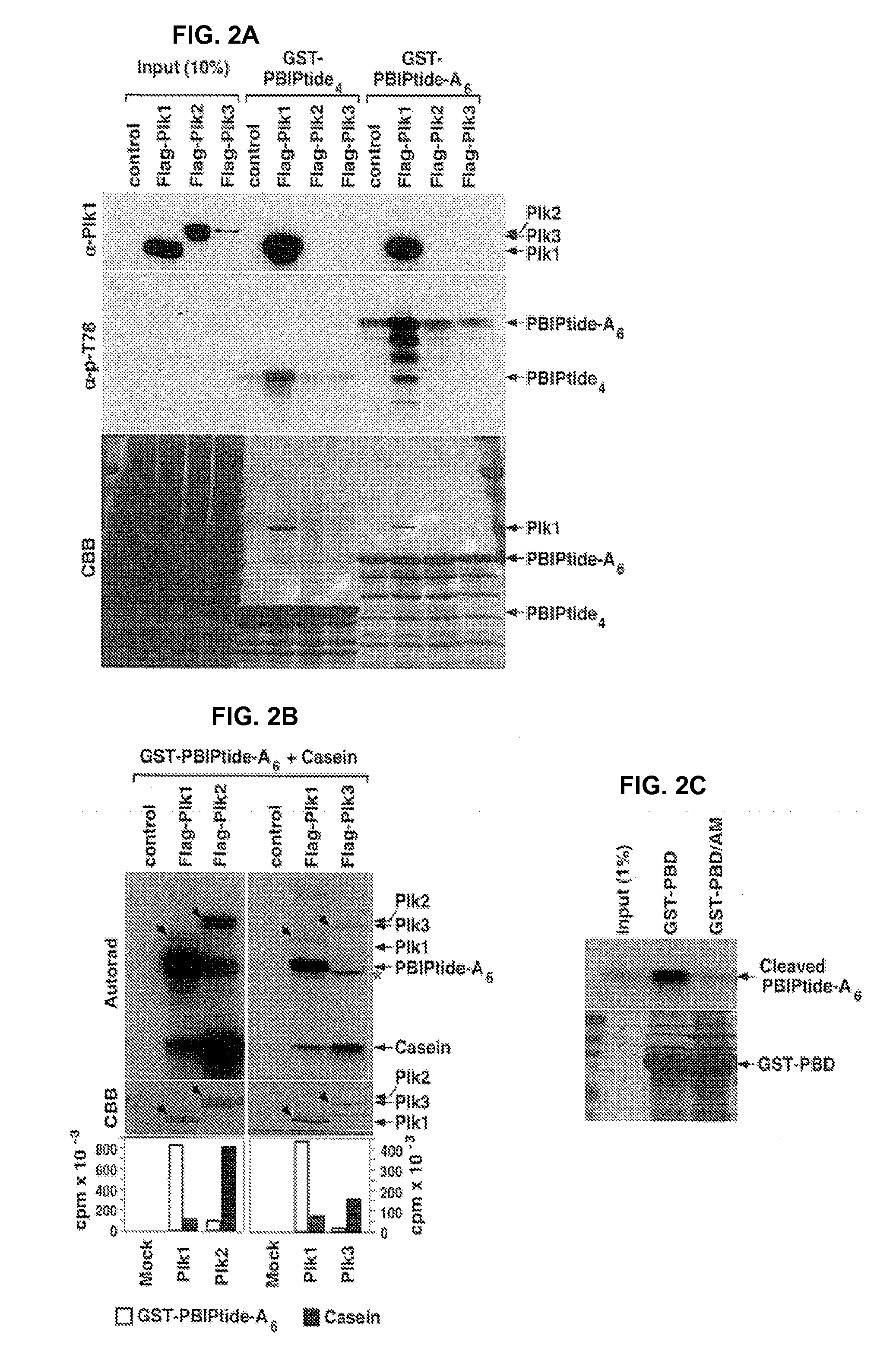
[0175]As shown in FIG. 2A-2C, Plk1, but not Plk2 or Plk3, phosphorylates and binds to the T78 motif of GST-PBIPtides. HeLa cell lysates obtained from HeLa cells expressing either Flag-Plk1, Flag-Plk2, or Flag-Plk3 were prepared in KC-plus buffer, and then incubated with the GST-PBIPtides (indicated in FIG. 2A-2C) immobilized on glutathione (GSH) agarose beads. The GST-PBIPtide precipitates were separated by SDS-PAGE, transferred to a PVDF membrane, and then immunoblotted with anti-Plk1 and anti-phospho-T78 antibody (see FIG. 2A). Afterward, the same membrane was stained with Coomassie (CBB) (see FIG. 2A). Note that the lysates expressing Plk1 efficiently generated the phospho-T78 epitope on GST-PBIPtides and, as a result, bound Plk1. The low levels of the phospho-T78 signals from the Plk2 or Plk3 transfected cells are likely due to the endogenous Plk...
PUM
| Property | Measurement | Unit |
|---|---|---|
| absorbance wavelength | aaaaa | aaaaa |
| absorbance wavelength | aaaaa | aaaaa |
| absorbance wavelength | aaaaa | aaaaa |
Abstract
Description
Claims
Application Information
 - R&D
- Intellectual Property
- Life Sciences
- Materials
- Tech Scout
- Unparalleled Data Quality
- Higher Quality Content
- 60% Fewer Hallucinations
Browse by: Latest US Patents, China's latest patents, Technical Efficacy Thesaurus, Application Domain, Technology Topic, Popular Technical Reports.
© 2025 PatSnap. All rights reserved.Legal|Privacy policy|Modern Slavery Act Transparency Statement|Sitemap|About US| Contact US: help@patsnap.com


