Light Activated Composite Tissue Adhesives

a composite tissue and light-activated technology, applied in the field of light-activated composite tissue adhesives, can solve the problems of high level of surgeon skill, inability to achieve clinical acceptance of methods, strength, toxicity and resorbability of tissue, etc., to achieve the effect of enhancing the solubility of chromophore, preventing chromophore, and increasing the solubility of collagen layer

Inactive Publication Date: 2011-05-26
CONVERSION ENERGY ENTERPRISES
View PDF10 Cites 21 Cited by
  • Summary
  • Abstract
  • Description
  • Claims
  • Application Information

AI Technical Summary

Benefits of technology

[0010]Consistent with an aspect of the present invention, a method of sealing and closing tissue and wounds is disclosed which comprises a composition that includes a derivatized collagen layer with incorporated photosensitive chromophores including riboflavin, lumichrome or lumiflavin and placed on the tissue. In another aspect of this invention the collagen composition is reacted with a solution of chromophore. Once the chromophore is uniformly distributed within the collagen composition, the layer or thin film is removed from solution and dried to remove any excess chromophore. Alternatively, the chromophore may include lumichrome or lumiflavin. In addition, the layer or film may be exposed to optical energy or light having a wavelength in a range of 360-375 nm (UVA) or 440-480 nm (blue light). Power densities may range from 0.1 W / cm2 to 2 W / cm2. The derivatized collagen can participate in the sealing and closure of wounds associated with trauma or surgery. ROS may be generated by the photo-degradation of the flavin and may disrupt the three-dimensional structure of collagen fibers found in tissues as well as in the derivatized collagen (flavin carrier) promoting crosslinking between tissue and the glue composite, improving both adhesive and cohesive strength.
[0012]Consistent with the present disclosure, several benefits may be provided as compared to the methods currently used for supplying reagents to tissues for sealing and bonding including solutions or gels that are applied to the wound area. The derivatized collagen incorporated chromophore layer can be sized to any shape or thickness and conforms to curved, flat or irregular tissue surfaces. The collagen compositions can be varied as well as the concentration of the chromophore to optimize the time release of the photosensitizing agent to the target site. The solubility of the chromophore is enhanced in the process of incorporation within the collagen composition. Other advantages include preventing the chromophore from migrating or diffusing to adjacent healthy tissue. The light is so configured so that it impinges only on the conformal layer. The physiological fluids of the tissue increase the solubility of the collagen layer resulting in the chromophore being released and diffused into the treatment area. The collagen layer may also help to obliterate wound dead spaces, and may be a useful adjunctive measure that could reduce the need for drains. The collagen layer appears to bond to wound collagen and as a result will aid in the apposition of the wound surfaces thus minimizing seroma (fluid) accumulation, which is a potential source of infection as these can be primarily or secondarily contaminated. The collagen layer also provides a moist environment an affect that is known to encourage healing. Light penetration is controlled by selection of wavelength range in order to activate only the released chromophore at the wound site.

Problems solved by technology

Methods to date have not gained clinical acceptance.
The major reasons include the high level of surgeon skill that is required, the strength, toxicity and resorbability of the tissue adhesive, the potential for irreparable laser damage and cost of the laser system.

Method used


Image

  • Light Activated Composite Tissue Adhesives
  • Light Activated Composite Tissue Adhesives
  • Light Activated Composite Tissue Adhesives

Examples


Embodiment Construction

[0028]Reference will now be made in detail to exemplary embodiments of the disclosure, examples of which are illustrated in the accompanying drawings. Wherever possible, the same reference numbers will be used throughout the drawings to refer to the same or like parts.

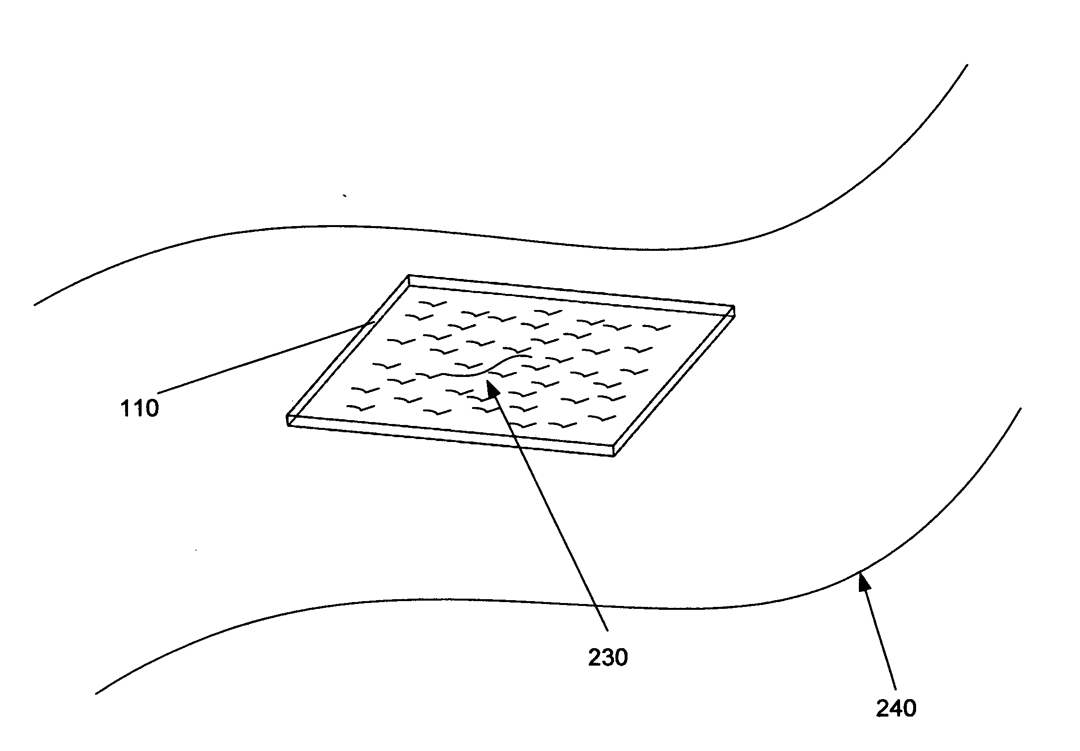
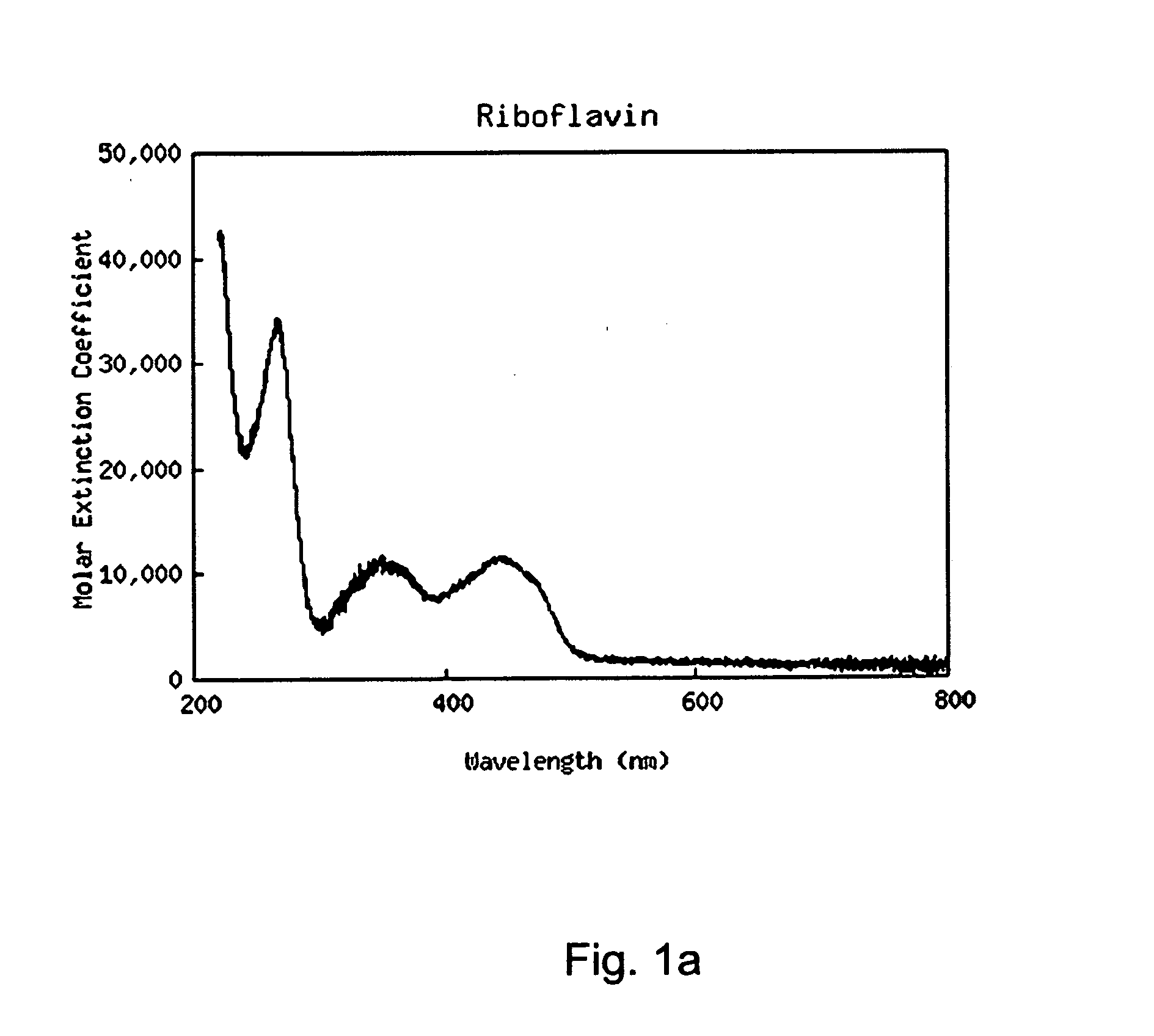
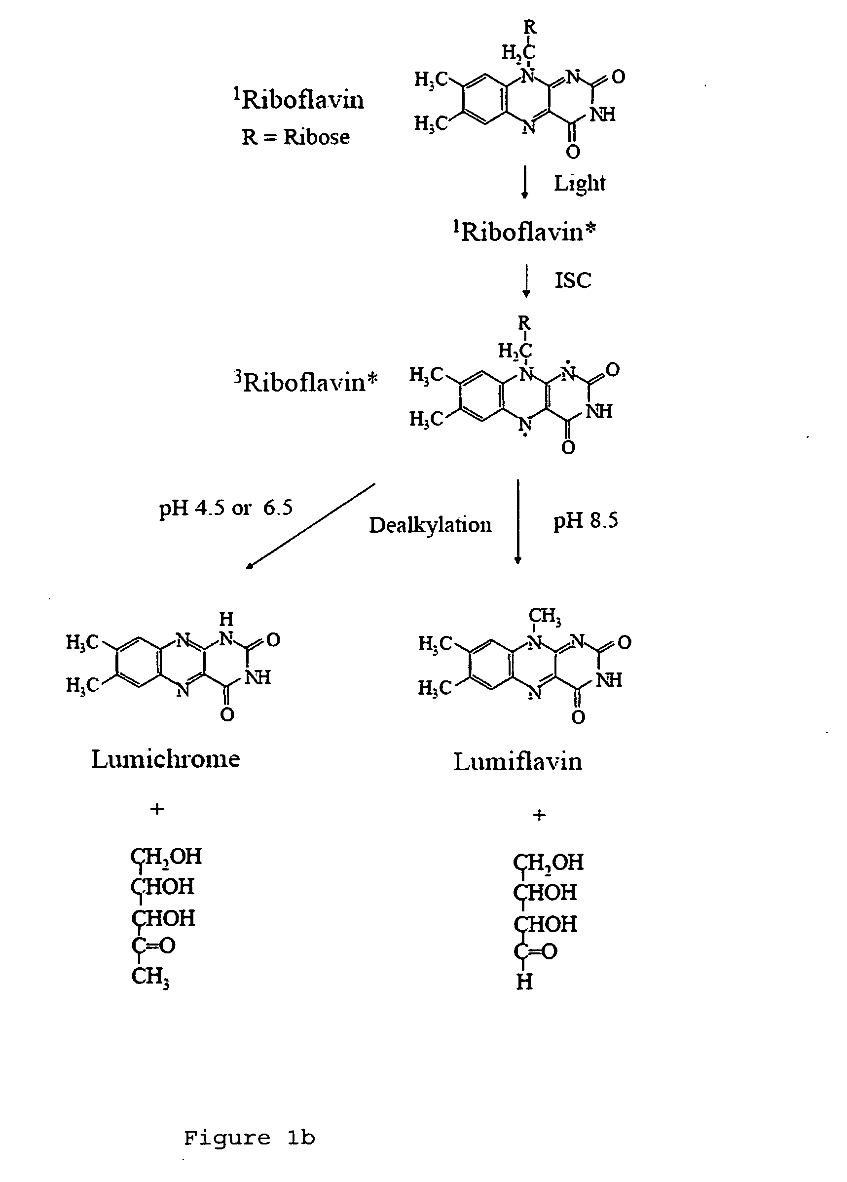
[0029]A layer of collagen that incorporates a photosensitive chromophore such as riboflavin, lumiflavin, lumichrome or a combination of thereof, for example, may be applied directly to a surgical incision or a trauma wound, for example. While the riboflavin diffuses to the affected area, the collagen layer is exposed to light (e.g., light having a wavelength between 360-375 nm or alternatively 440-480 nm) with power densities in the range 0.1 W / cm2 to 2 W / cm2. In other examples, the collagen layer may instead contain lumichrome or lumiflavin. A method of preparing a composition consistent with the present disclosure will next be described.

[0030]The collagen layer may be made from a collagen which has been extracted, pu...


PUM

PropertyMeasurementUnit
concentrationaaaaaaaaaa
wavelengthaaaaaaaaaa

Abstract

A light activated collagen-flavin composite layer incorporating riboflavin is applied as treatment for infected lesions caused by bacteria and as the consequence of surgical procedures. These composites have also been found to be strong tissue adhesives that are effective in closing and sealing wounds, fixation of grafts / implants and anastomoses. Advantages include speed of closure, reduced infection due to the elimination of foreign matter, evidence of accelerated wound healing and the ease of use in complex surgery, especially when watertight seals, limited access or small repair size are important factors. The riboflavin in the collagen layer is exposed to light (e.g., light having a wavelength between 360-375 nm or 440-480 nm), decomposing the riboflavin to form reactive oxygen species (ROS). Strong crosslinks between the collagen composite and tissue results. In addition, similar exposures eradicate pathogens in the wound is eradicated resulting in a sterile wound. In other examples, the composite film may instead contain lumichrome or lumiflavin.

Description

[0001]The present application claims the benefit of U.S. Provisional Patent Application No. 61 / 280,925, filed Nov. 10, 2010, the entire contents of which are incorporated herein by reference.I. BACKGROUND[0002]There has been heightened interest in developing light activated tissue solders and sealants as a substitute for conventional closure methods such as the fixation of grafts / implants and anastomoses. Advantages include speed of closure, reduced infection due to the elimination of foreign matter, evidence of accelerated wound healing and the ease of use in complex surgery, especially when watertight seals, limited access or small repair size are important factors. Laser activation provides a directed energy source for precise placement of the sealing material and can amplify the crosslinking mechanism for bonding together incisional edges or wound tissue flaps.[0003]Numerous studies have reported the efficacy of light activated materials to join soft tissues. The availability of...

Claims


Application Information

Patent Timeline
Patent Type & Authority Applications(United States)
IPC IPC(8): A61B17/00A61K38/39A61K9/00A61P17/02
CPCA61B17/00491A61B2017/005A61K9/7007A61K31/525A61K38/39A61K47/42A61L31/14A61L31/044A61K2300/00A61P17/02A61P43/00A61B2017/00004A61B2017/00517A61B2017/00884A61B2017/00951A61L24/0015A61L24/102A61L2300/428
Inventor SOLTZ, BARBARA A.SOLTZ, ROBERT
Owner CONVERSION ENERGY ENTERPRISES
Why Eureka
Try Eureka