
Providing an ultrasound spatial compound image in an ultrasound system

a technology of ultrasound system and compound image, applied in the field of ultrasound system, can solve the problem of difficult to precisely comprehend the correlation between the 2d ultrasound image and the 3d ultrasound imag

Active Publication Date: 2011-06-09
SAMSUNG MEDISON
View PDF5 Cites 5 Cited by
  • Summary
  • Abstract
  • Description
  • Claims
  • Application Information

AI Technical Summary

Benefits of technology

The Summary is provided to introduce a selection of concepts in a simplified form that are further described below in the Detailed Description. This Summary is ...

Problems solved by technology

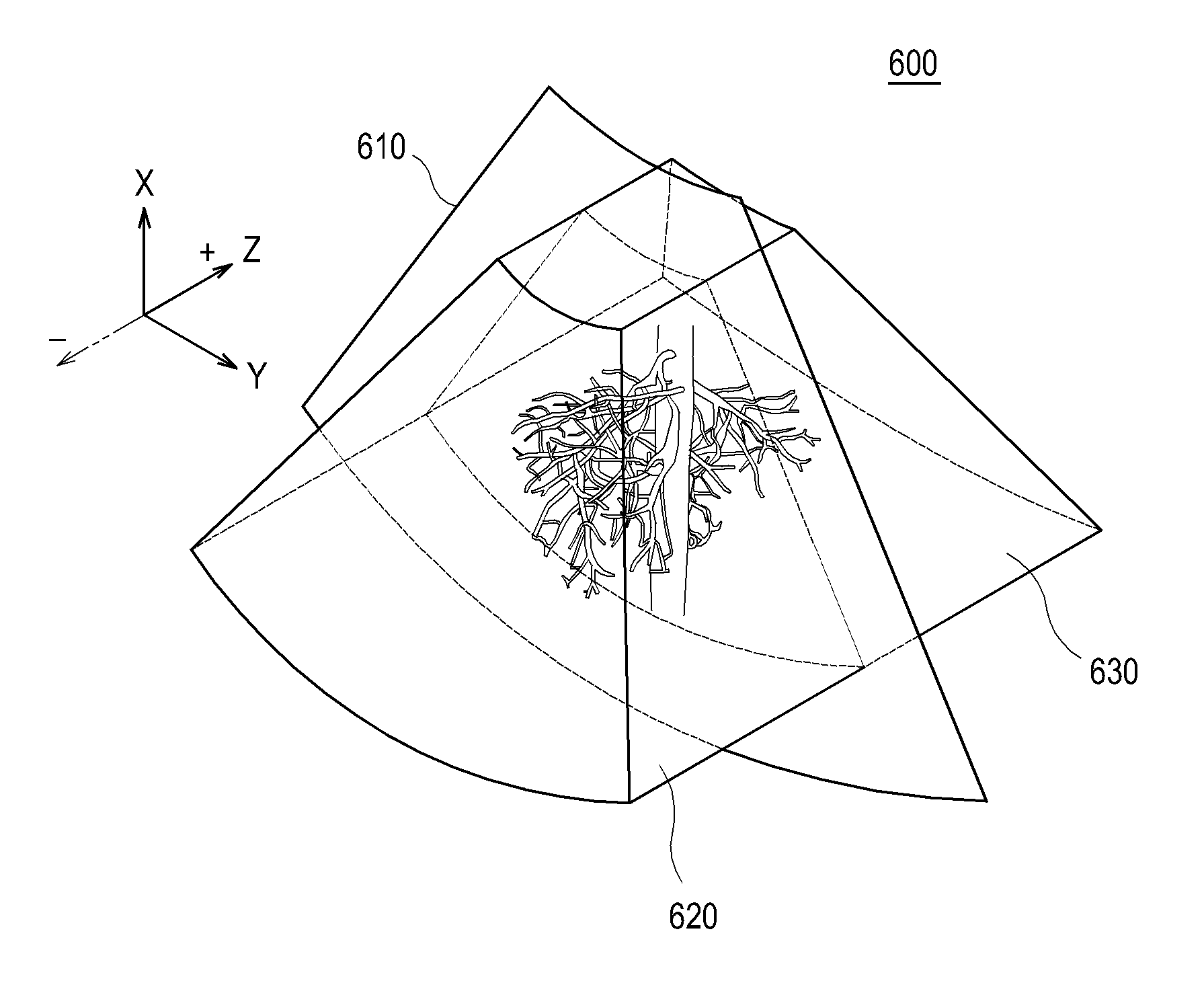
However, it may be difficult to precisely comprehend a correlation between the 2D ultrasound image and the 3D ultrasound image.

Method used


Image

  • Providing an ultrasound spatial compound image in an ultrasound system
  • Providing an ultrasound spatial compound image in an ultrasound system
  • Providing an ultrasound spatial compound image in an ultrasound system

Examples


Embodiment Construction

A detailed description may be provided with reference to the accompanying drawings. One of ordinary skill in the art may realize that the following description is illustrative only and is not in any way limiting. Other embodiments of the present invention may readily suggest themselves to such skilled persons having the benefit of this disclosure.

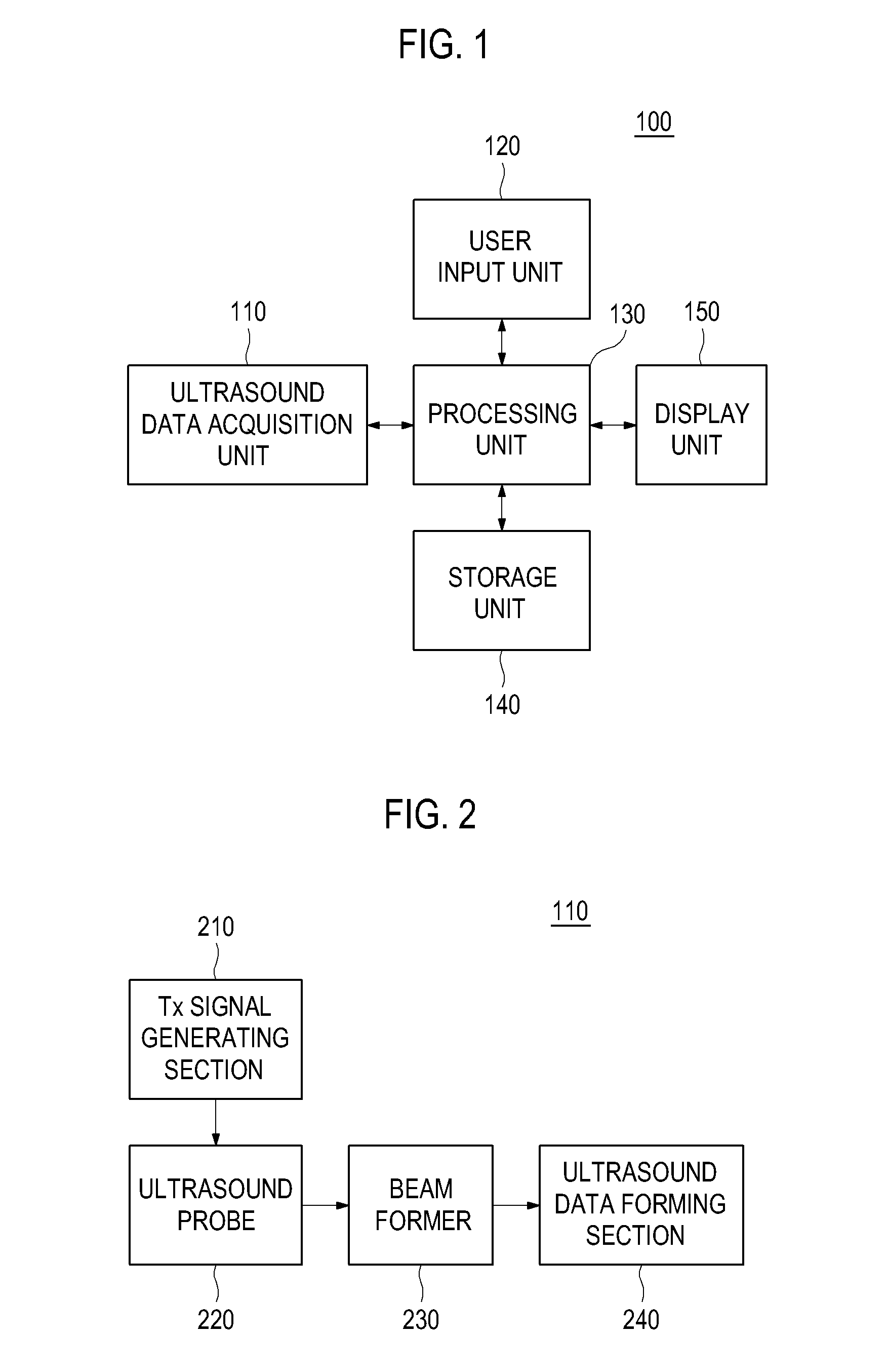
Referring to FIG. 1, an ultrasound system 100 in accordance with an illustrative embodiment is shown. As depicted therein, the ultrasound system 100 may include an ultrasound data acquisition unit 110. The ultrasound data acquisition unit 110 may be configured to transmit and receive ultrasound signals to and from a target object to output ultrasound data.

FIG. 2 is a block diagram showing an illustrative embodiment of the ultrasound data acquisition unit 110. Referring to FIG. 2, the ultrasound data acquisition unit 110 may include a transmit (Tx) signal generating section 210, an ultrasound probe 220, a beam former 230 and an ultrasound da...


PUM


Abstract

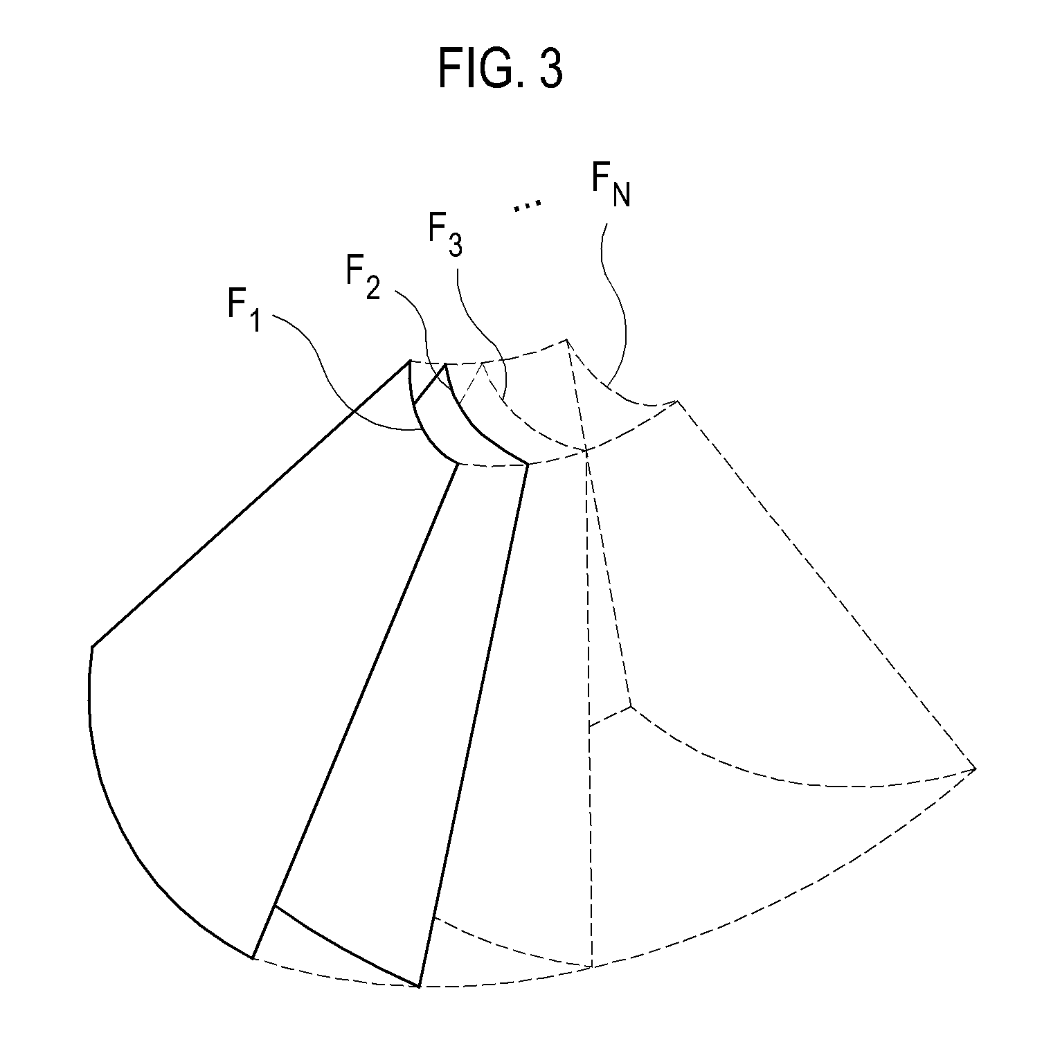
Embodiments for providing an ultrasound spatial compound image are disclosed. In one embodiment, by way of non-limiting example, an ultrasound system comprises: an ultrasound data acquisition unit configured to transmit and receive ultrasound signals to and from a target object to output first ultrasound data and a plurality of sets of second ultrasound data corresponding to a region of interest (ROI); a user input unit configured to receive input information for defining the ROI; and a processing unit in communication with the ultrasound data acquisition unit and the user input unit, the processing unit being configured to form volume data based on the plurality of sets of second ultrasound data, compare the first ultrasound data with the volume data to detect geometric information therein, form a two-dimensional (2D) ultrasound image based on the first ultrasound data, and a first three-dimensional (3D) ultrasound image and a second 3D ultrasound data based on the volume data in consideration of the geometric information, and perform a spatial compound upon the 2D ultrasound image, the first 3D ultrasound image and the second 3D ultrasound image based on the geometric information to form an ultrasound spatial compound image.

Description

CROSS-REFERENCE TO RELATED APPLICATIONSThe present application claims priority from Korean Patent Application No. 10-2009-0121590 filed on Dec. 9, 2009, the entire subject matter of which is incorporated herein by reference.TECHNICAL FIELDThe present disclosure generally relates to ultrasound systems, and more particularly to providing an ultrasound spatial compound image of two-dimensional and three-dimensional ultrasound images in an ultrasound system.BACKGROUNDAn ultrasound system has become an important and popular diagnostic tool since it has a wide range of applications. Specifically, due to its non-invasive and non-destructive nature, the ultrasound system has been extensively used in the medical profession. Modern high-performance ultrasound systems and techniques are commonly used to produce two-dimensional (2D) or three-dimensional (3D) ultrasound images of internal features of an object (e.g., human organs).The ultrasound system may provide the 3D ultrasound image includi...

Claims


Application Information

Patent Timeline
IPC IPC(8): A61B8/14
CPCG01S15/8993G01S7/52063G01S7/52071G01S7/52074G06T7/0012A61B8/5215A61B8/13A61B8/085A61B8/5253A61B8/469A61B8/14G06T11/00G06T17/00
Inventor LEE, SUK JINKIM, JUNG
Owner SAMSUNG MEDISON
Learn More