Methods for structural analysis of glycans
- Summary
- Abstract
- Description
- Claims
- Application Information
AI Technical Summary
Benefits of technology
Problems solved by technology
Method used
Image

Examples
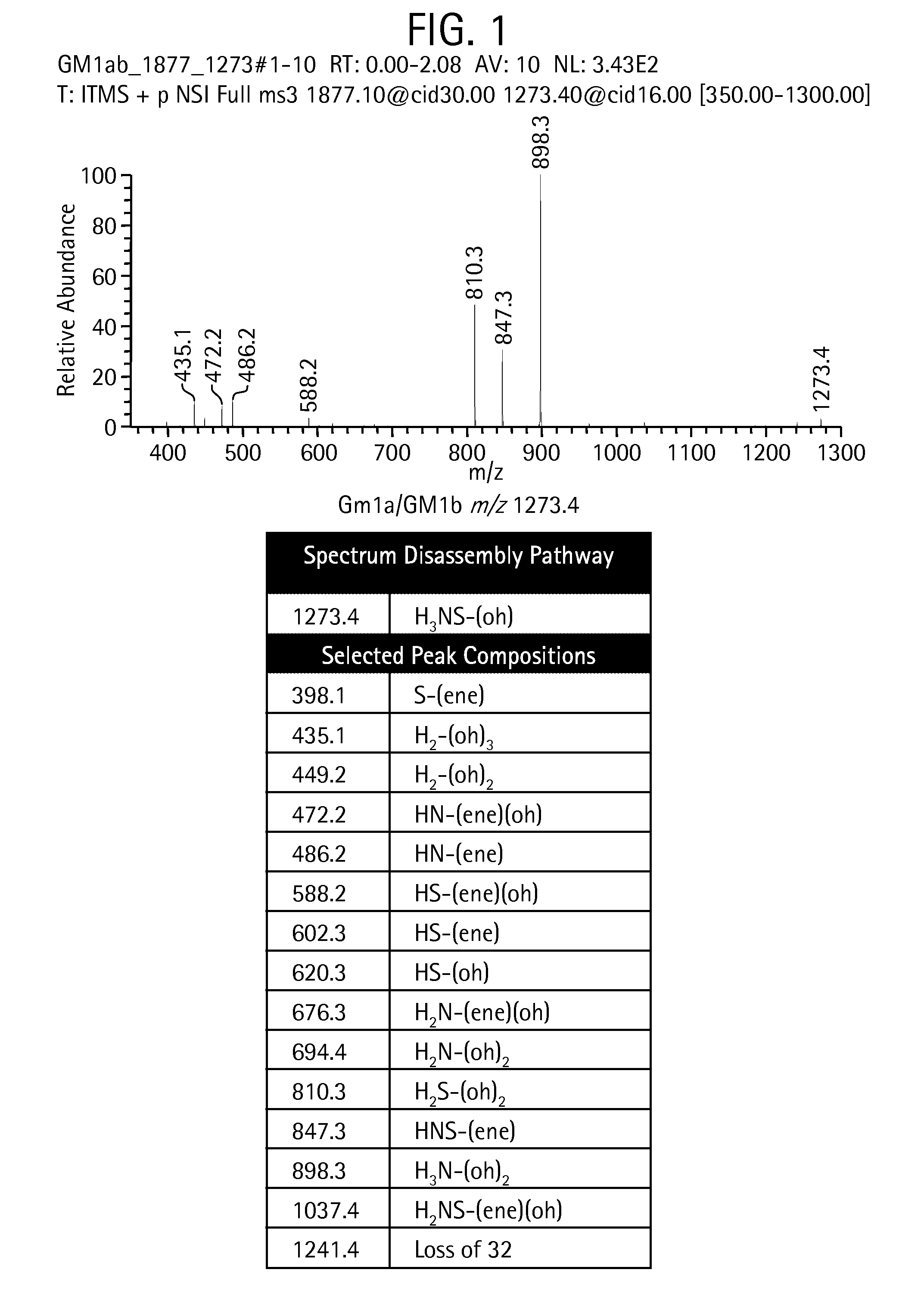
example 1
Fragmentation of Permethylated Glycans in Positive Experimental Mode
[0299]The below data show that some chemical bonds in permethylated glycans are considerably more likely to rupture (i.e., these bonds are more “labile”) than others, and therefore lead to predicable fragments when the glycans are analyzed via MSn.
[0300]It has been well established that permethylated glycans tend to fragment most readily at the glycosidic bonds between residues, especially when the number of residues in the precursor fragment is, for example, four or more. A closer examination shows that certain permethylated residues form weaker glycosidic bonds, leading to a skewed distribution of fragment intensities on the experimental spectrum. That is, fragments formed by the rupture of weak bonds tend to occur with a higher relative abundance than fragments formed by the rupture of strong bonds.
[0301]Metal ion (Na+, K+, and Li+) and proton localization (or charge localization) in positive mode and electron de...
example 2
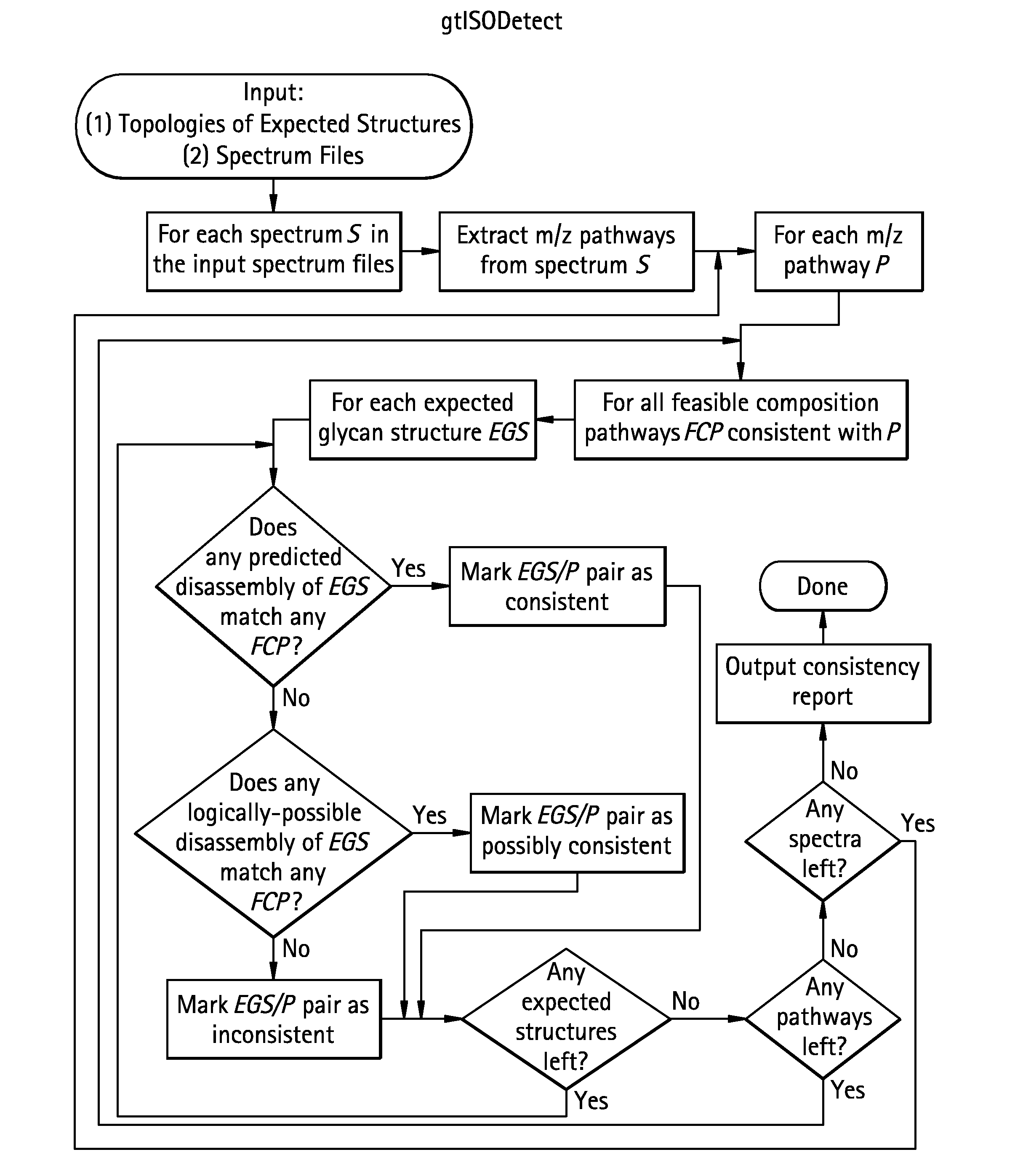
The gtIsoDetect Algorithm Applied to Ovalbumin m / z 1677.8
[0325]
[0326]To illustrate the gtIsoDetect algorithm, we apply it to the concrete example of two isomeric glycans found in ovalbumin m / z 1677.8. The composition pathway used in this example are shown in FIG. 4 and the two isomeric structures under consideration—labeled B and C in accordance with Ashline et al, Anal Chem 79: 3830-3842 (2007)—are shown in Scheme 12.
[0327]Processing 1677.8→1384.5→1125.4→866.4→662.4→444.1
[0328]First we demonstrate how gtIsoDetect applies the m / z pathway 1677.8→1384.5→1125.4→866.4→662.4→444.1 to structures B and C. For both structures in parallel, substructures are sought that match the composition of each successive ion in the pathway as shown in Table 9.
TABLE 9Substructure Embed-Substructure Embed-m / zCompositionded in Structure Bded in Structure C1677.8H3N3nH1H2H3N4N5N6n7H1H2H3N4N5N6n71384.5H3N3-(ene)H1H2H3N4N5N6H1H2H3N4N5N61125.4H3N2-(ene)(oh)H1H2H3N5N6 ORH1H2H3N5N6 ORH1H2H3N4N6H1H2H3N4N6866.4H3N...
example 3
gtSequenceGrow for Glycan Sequencing
[0340]In this Example, we use the gtSequenceGrow method to assign a glycan topology. These data were collected via MSn, but this technique can be applied to any technology that fragments glycans in a predictable step-wise manner such as, for example, with a series of glycosidase digests interleaved with MS / MS analysis.
[0341]Processing follows the chart of FIG. 3. We begin processing at the terminal spectrum m / z 458.1. The example is slightly simplified in that m / z 1677.7 has two possible compositions—H3N3n or H2N4h—but we exclude the second possibility because the MS3 spectrum (m / z 13384.5) is consistent with only the first. gtSequenceGrow is applied according to the following manner:
Simulate m / z 458.1 / HN-(ene)(oh)2
[0342]Create all substructure matching compositions without scars (Scheme 13).
Scheme 13
[0343]1) H—N[0344]2) N—H[0345]Add all combinations of scars. (Scheme 14).
[0346]The structure numbering scheme is according to the following guidelin...
PUM
| Property | Measurement | Unit |
|---|---|---|
| Fraction | aaaaa | aaaaa |
| Fraction | aaaaa | aaaaa |
| Fraction | aaaaa | aaaaa |
Abstract
Description
Claims
Application Information