Endoscope having a shaft tube and optic

a technology of endoscope and shaft tube, which is applied in the field of surgical endoscopes, can solve the problems of having to manoeuvre and follow up the extracted liquid, and achieve the effect of reducing the negative impact on the patient and facilitating cleaning for reus

Inactive Publication Date: 2011-07-07
OLYMPUS WINTER & IBE
View PDF8 Cites 38 Cited by
  • Summary
  • Abstract
  • Description
  • Claims
  • Application Information

AI Technical Summary

Benefits of technology

[0014]Advantageously, according to claim 3, the optic and the morcellator can be removed from the shaft tube. This facilitates cleaning for reuse and creates opportunities for combination with other instruments.
[0015]Advantageously according to claim 4, the shaft tube is formed in such a way that it can also be used as the outer shaft of a resection device mentioned at the outset. This creates the great advantage that the same outer shaft can be used for the resection and for subsequent morcellating. Thus, it can remain in situ in the urethra throughout the operation, therefore reducing the negative impact on the patient from inserting and removing the shaft several times.

Problems solved by technology

Because poor visibility prevails during such operations, on account of bleeding, thorough flushing is essential.
Similar problems exist with other surgical extraction instruments, such as with simple suction tubes, with which, for example, tissue residues, stone fragments or the like are sucked up from the base of the bladder.
Here too, as with the morcellating work described above, we are faced with the problem of having to manoeuvre the suction tube or the suction morcellator with the suction aperture under targeted optic observation and of having to follow up the extracted liquid.

Method used


Image

  • Endoscope having a shaft tube and optic
  • Endoscope having a shaft tube and optic

Examples


Embodiment Construction

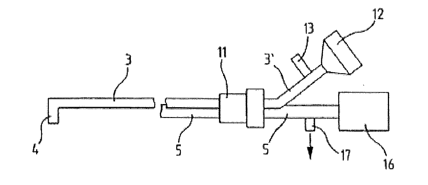
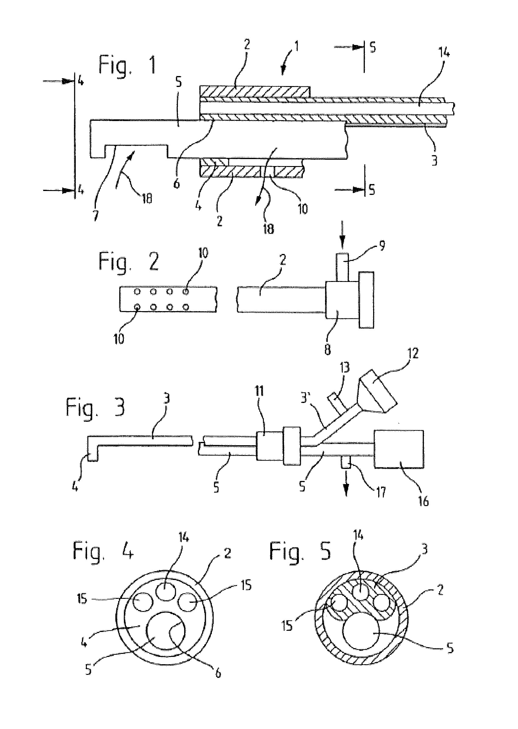
[0022]FIG. 1 shows, in longitudinal section, the distal tip of a urological endoscope 1 with a shaft tube 2 forming the outer wall and an optic 3 located therein, which, as FIGS. 1 and 3 show, is formed in the distal end region as a sealing element 4, which seals the internal cross-section of the shaft tube 2. Proximally adjacent to the sealing element 4, however, as FIG. 1 shows along the section line 5-5, the cross-section of the optic 3 is smaller, which is also quite clearly shown in FIG. 5.

[0023]In the free internal cross-section of the shaft tube 2 available at this point next to the optic 3, there is space to introduce an elongated, rod shaped morcellator 5, which, as shown in FIGS. 1 and 4, passes through a hole 6 parallel to the axis of the shaft tube 2, in the sealing element 4 and is positioned with its distal end area in front of the distal end of the shaft tube 2. Here, the morcellator 5 has a side suction aperture 7, which is used for morcellating and sucking up the mo...


PUM


Abstract

A surgical endoscope having a shaft tube that can be connected at its proximal end area to a flushing water inlet and in the distal end area, has at least one lateral opening and having an optic extending through the shaft tube, said optic being designed with a smaller cross-section than the internal cross-section of the shaft tube, and through said optic an image guide extends along the length thereof, where a suction tube is arranged in the free internal cross-section of the shaft tube next to the optic, the suction opening of said suction tube being arranged distally in front of the shaft tube, said suction tube also being proximally connected to a suction device, and where in the distal end region of the shaft tube a locking element, locking the remaining free internal cross-section, thereof is disposed.

Description

CROSS-REFERENCE TO RELATED APPLICATION[0001]The present application is based upon and claims the benefit of priority from the PCT / EP2009 / 004499 filed on Jun. 23, 2009, the entire contents of which are incorporated herein by reference.BACKGROUND[0002]1. Field[0003]The present invention generally relates to surgical endoscopes, and particularly to a surgical endoscope having a shaft tube and an optic located within it, with which the area in front of the distal end of the shaft tube can be observed.[0004]2. Description of the Related Art[0005]The use of endoscopes for prostate resection is known. They comprise a blade arranged in the shaft tube, which can cut off tissue in front of the distal end of the shaft tube. Normally this blade is developed as an HF-cutting loop. Because poor visibility prevails during such operations, on account of bleeding, thorough flushing is essential.[0006]Such endoscopes used for resection comprise a continuous flushing system, where the visible area is ...

Claims


Application Information

Patent Timeline
Patent Type & Authority Applications(United States)
IPC IPC(8): A61B1/012
CPCA61B1/00094A61B1/00135A61B1/0014A61B2017/320775A61B1/12A61B1/307A61B2017/22079A61B1/012
Inventor REIF, MATTHIASWOSNITZA, THOMAS
Owner OLYMPUS WINTER & IBE
Why Eureka
Try Eureka